. 2016 Jul 26:7:1081.
doi: 10.3389/fpls.2016.01081. eCollection 2016.

The Root Hair Specific SYP123 Regulates the Localization of Cell Wall Components and Contributes to Rizhobacterial Priming of Induced Systemic Resistance

Affiliations

The Root Hair Specific SYP123 Regulates the Localization of Cell Wall Components and Contributes to Rizhobacterial Priming of Induced Systemic Resistance

Cecilia Rodriguez-Furlán et al. Front Plant Sci. .

Abstract

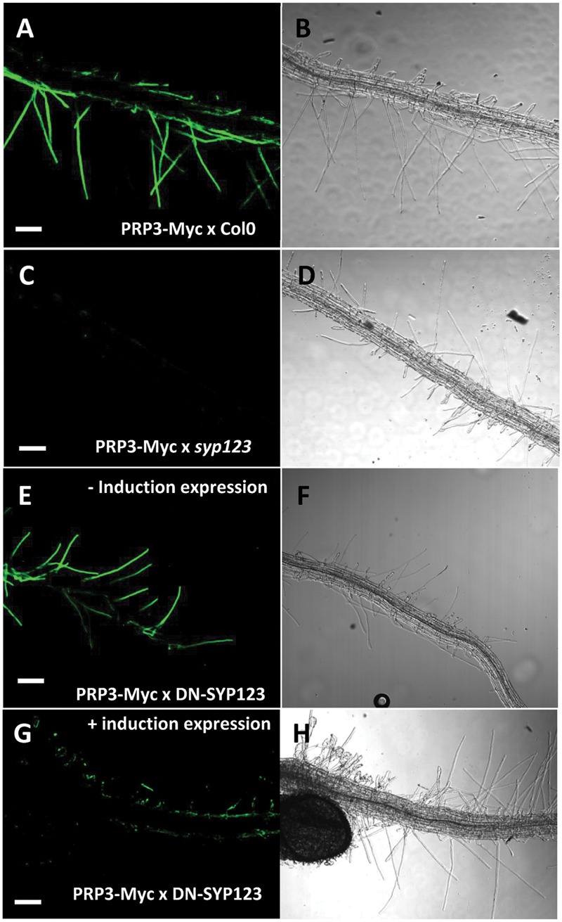
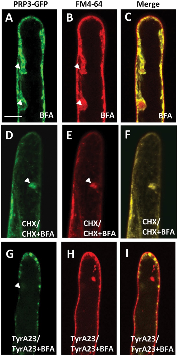
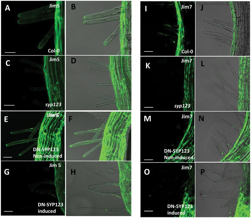
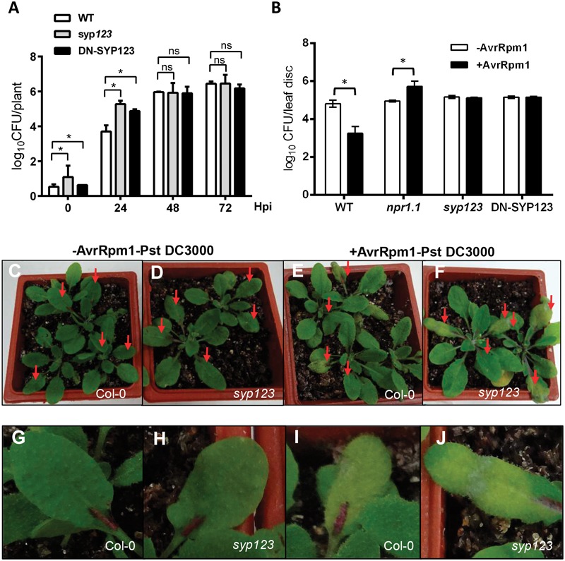
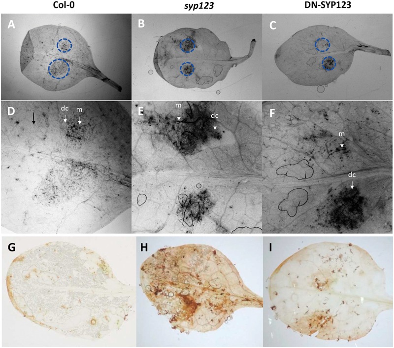
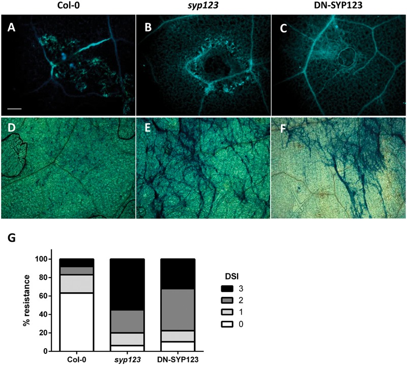
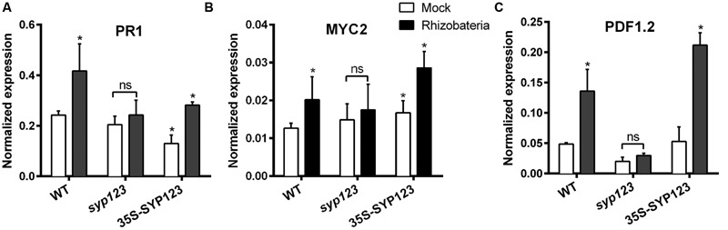
Root hairs are important for nutrient and water uptake and are also critically involved the interaction with soil inhabiting microbiota. Root hairs are tubular-shaped outgrowths that emerge from trichoblasts. This polarized elongation is maintained and regulated by a robust mechanism involving the endomembrane secretory and endocytic system. Members of the syntaxin family of SNAREs (soluble N-ethylmaleimide-sensitive factor attachment protein receptor) in plants (SYP), have been implicated in regulation of the fusion of vesicles with the target membranes in both exocytic and endocytic pathways. One member of this family, SYP123, is expressed specifically in the root hairs and accumulated in the growing tip region. This study shows evidence of the SYP123 role in polarized trafficking using knockout insertional mutant plants. We were able to observe defects in the deposition of cell wall proline rich protein PRP3 and cell wall polysaccharides. In a complementary strategy, similar results were obtained using a plant expressing a dominant negative soluble version of SYP123 (SP2 fragment) lacking the transmembrane domain. The evidence presented indicates that SYP123 is also regulating PRP3 protein distribution by recycling by endocytosis. We also present evidence that indicates that SYP123 is necessary for the response of roots to plant growth promoting rhizobacterium (PGPR) in order to trigger trigger induced systemic response (ISR). Plants with a defective SYP123 function were unable to mount a systemic acquired resistance in response to bacterial pathogen infection and ISR upon interaction with rhizobacteria. These results indicated that SYP123 was involved in the polarized localization of protein and polysaccharides in growing root hairs and that this activity also contributed to the establishment of effective plant defense responses. Root hairs represent very plastic structures were many biotic and abiotic factors can affect the number, anatomy and physiology of root hairs. Here, we presented evidence that indicates that interactions with soil PGPR could be closely regulated by signaling involving secretory and/or endocytic trafficking at the root hair tip as a quick way to response to changing environmental conditions.

Keywords: PRP3; cell wall; induced systemic resistance; plant growth promoting rhizobacterium; rhizobacteria; syntaxin; systemic acquired resistance; trafficking.


Figures

FIGURE 1
FIGURE 1
SYP123 defective plants have altered PRP3 distribution at the root hair cell wall. PRP3-Myc protein immunolocalization without permeabilization in roots of Arabidopsis seedlings in the Col-0 (A,B), syp123 (C,D), the dominant negative (DN)-SYP123 with induction of the expression (E,F), and the control without induction (G,H). Immunolocalization was performed using Anti-Myc mouse monoclonal primary antibody and Alexa Fluor-488 conjugated goat anti-mouse IgG (green fluorescence left panel). Bright field images are displayed on the right panel. Scale bar = 50 μm.
FIGURE 2
FIGURE 2
Brefeldin A (BFA) treatment caused the internalization of PRP3-GFP. PRP3-GFP (A) and FM4-64 (B) are present in BFA compartments, evidenced by the co-localization in the merged image (C). Cycloheximide treatment inhibited protein synthesis but did not inhibit the internalization of PRP3-GFP (D) or FM4-64 (E) into BFA compartments as they still co-localize (F). TyrA23 treatments efficiently inhibited BFA-induced intracellular accumulation of PRP3-GFP (G) and FM4-64 (H). Merged image (I). Scale bar = 10 μm.
FIGURE 3
FIGURE 3
SYP123 defective plants exhibit altered distribution of JIM5 epitopes. Immunodetection in 5-days-old roots using the JIM5 antibody (A–H), which detects de-esterified pectins, or JIM7 (I–P), which detects esterified pectins. The Jim5 signal was strong in the root hair of Col-0 wild-type (WT) plants, and Jim7 signal was absent in the tips of syp123 and in the DN (induced with dexamethasone and heat). Scale bars are indicated in the figure. Scale bar = 20 μm.
FIGURE 4
FIGURE 4
syp123 mutants are susceptible to Pseudomonas syringae infection. (A) Leaves of the indicated genotypes were infiltrated with Pst DC3000 and the number of colony forming units (CFUs) per leaf area determined after serial dilution and cultivation on selective media. Mean and standard deviation of triplicates at 0, 2, and 5 days post-infection (dpi) are shown. Different letters represent statistical differences during the progress of the infection (non-significant differences between genotypes were recorded with ANOVA and Tukey’s post-test). (B) Whole leaves of 4-weeks-old, soil-grown, WT Col-0 and mutant plants were infiltrated with Pst AvrRpm1 to trigger SAR (+AvrRpm1); a solution of 10 mM MgCl2 was used as mock (-AvrRpm1). After 24 h the systemic leaves were infiltrated with Pst DC3000. Bacterial growth was monitored 72-h post-infection. Error bars represent standard deviation from six samples. Different letters represent statistical differences between the genotypes and treatment at p < 0.01 (Tukey’s test). All experiments were performed in triplicate with similar results. (C–F) Pictures of WT and syp123 mutant plants showing the phenotype 72-h post-infection. (G–J) A Close up of lesions in infected leaves.
FIGURE 5
FIGURE 5
Botrytis infection assays in seedlings with a defective SYP123 function. (A–F) The leaves from Col-0, syp123 mutant, and DN-SYP123 plants were inoculated with a spore suspension of Botrytis cinerea. Leaves were stained with trypan blue at 4 days post inoculation (dpi). Dotted circles indicate the borders of spore inoculation sites in control Col-0 (A,D), syp123 mutant (B,E), and DN-SYP123 (C,F). Stained plant cells surrounding the fungal mycelium (m) indicates dead plant cells (dc). (D–F) Shows a higher magnification of the inset from the superior panel at 4 dpi. (G–I) Production of H2O2 is visualized by DAB staining in Botrytis inoculated leaves at 4 dpi. The brown precipitate shows DAB polymerization at the site of H2O2 production. Representative leaves detached from inoculated Col-0 (A), syp123, and DN-SYP123 plants are shown. All the experiments were performed in triplicate with similar results.
FIGURE 6
FIGURE 6
Phytophthora infection assays in seedlings with a defective SYP123 function. (A–F) Response to Phytophthora infestans inoculation. Callose staining with aniline blue in Arabidopsis WT Col-0 (A), syp123 mutant (B), and DN-SYP123 (C) 7 dpi. Callose accumulated at the site of inoculation in all plants. A small amount of callose was deposited in Col-0 (A), while widespread callose deposition occurred in syp123 (B) and DN-SYP123 (C) leaves. Trypan blue staining of the inoculated leaves of Col-0 (D), syp123 (E), and DN-SYP123 (F) at 7 dpi. Scale Bar = 50 μm. (G) Disease severity index (DSI) presented by numbers from “0” to “3”, as follows: 0 = no disease symptoms; 1 = a few spots observed within the droplet/plug zone; 2 = confined lesion covering droplet/plug zone; 3 = outgrowing lesion. Mean disease resistance scores were transformed into percentage values for comparison of replicate inoculations.
FIGURE 7
FIGURE 7
Induced systemic response (ISR)-marker genes expression is affected by SYP123 levels. The mRNA levels of (A) PR1 (B) MYC2, and (C) PDF1.2 ISR marker genes were measured in the rosette leaves of WT Col-0, syp123, or syp123x35S:SYP123 plants. The plants were grown for 7 days and then the roots were inoculated with beneficial rhizobacteria Pseudomonas spp. At 8 dpi the leaves were collected and the expression of ISR marker genes was analyzed using qRT-PCR. Asterisks indicate statistically significant differences (Student’s t-test, p < 0.01) compared to the control.

References

    1. Adam L., Somerville S. C. (1996). Genetic characterization of five powdery mildew disease resistance loci in Arabidopsis thaliana. Plant J. 9 341–356. 10.1046/j.1365-313X.1996.09030341.x - DOI - PubMed
    1. Albersheim P., Darvill A., Roberts K., Sederoff R., Staehelin A. (2011). “Biochemistry of the cell wall molecules,” in Plant Cell Walls: From Chemistry to Biology eds Albersheim P., Darvill A., Roberts K., Serderoff R., Staehelin A. (New York, NY: Garland Science; ) 67–118.
    1. Baluška F., Hlavacka A., Šamaj J., Palme K., Robinson D. G., Matoh T., et al. (2002). F-actin-dependent endocytosis of cell wall pectins in meristematic root cells. Insights from brefeldin a-induced compartments. Plant Physiol. 130 422–431. - PMC - PubMed
    1. Baluška F., Salaj J., Mathur J., Braun M., Jasper F., Šamaj J., et al. (2000). Root hair formation: f-actin-dependent tip growth is initiated by local assembly of profilin-supported F-actin meshworks accumulated within expansin-enriched bulges. Dev. Biol. 227 618–632. 10.1006/dbio.2000.9908 - DOI - PubMed
    1. Bellincampi D., Cervone F., Lionetti V. (2014). Plant cell wall dynamics and wall-related susceptibility in plant-pathogen interactions. Front. Plant Sci 5:228 10.3389/fpls.2014.00228 - DOI - PMC - PubMed

LinkOut - more resources