Review
. 2016 Jul 18:5:F1000 Faculty Rev-1735.
doi: 10.12688/f1000research.7864.1. eCollection 2016.

Transporting mitochondria in neurons

Affiliations
Review

Transporting mitochondria in neurons

Meredith M Course et al. F1000Res. .

Abstract

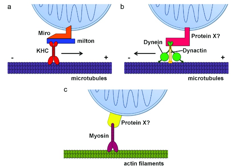
Neurons demand vast and vacillating supplies of energy. As the key contributors of this energy, as well as primary pools of calcium and signaling molecules, mitochondria must be where the neuron needs them, when the neuron needs them. The unique architecture and length of neurons, however, make them a complex system for mitochondria to navigate. To add to this difficulty, mitochondria are synthesized mainly in the soma, but must be transported as far as the distant terminals of the neuron. Similarly, damaged mitochondria-which can cause oxidative stress to the neuron-must fuse with healthy mitochondria to repair the damage, return all the way back to the soma for disposal, or be eliminated at the terminals. Increasing evidence suggests that the improper distribution of mitochondria in neurons can lead to neurodegenerative and neuropsychiatric disorders. Here, we will discuss the machinery and regulatory systems used to properly distribute mitochondria in neurons, and how this knowledge has been leveraged to better understand neurological dysfunction.

Keywords: Myosins; Transporting mitochondria; dynein; mitochondria; neurons.


Conflict of interest statement

Competing interests: The author(s) declared that they have no competing interests.

No competing interests were disclosed.

Figures

Figure 1.
Figure 1.. Schematic representation of mitochondrial transport machinery.
( a) The primary motor/adaptor complex mediating anterograde mitochondrial transport along microtubules (purple), including kinesin heavy chain (KHC) (red), Miro (orange), and milton (blue). ( b) The machinery mediating retrograde mitochondrial transport along microtubules (purple), including dynein (green), dynactin (gold), and a potential motor adaptor, Protein X (pink). Protein X could be the milton/Miro complex . ( c) Mitochondrial movement along actin filaments (olive), using a myosin motor (fuschia) and a potential motor adaptor, Protein X (yellow).

References

    1. Rossier MF: T channels and steroid biosynthesis: in search of a link with mitochondria. Cell Calcium. 2006;40(2):155–64. 10.1016/j.ceca.2006.04.020 - DOI - PubMed
    1. Oh-hama T: Evolutionary consideration on 5-aminolevulinate synthase in nature. Orig Life Evol Biosph. 1997;27(4):405–12. 10.1023/A:1006583601341 - DOI - PubMed
    1. Kugler P, Baier G: Mitochondrial enzymes related to glutamate and GABA metabolism in the hippocampus of young and aged rats: a quantitative histochemical study. Neurochem Res. 1992;17(2):179–85. 10.1007/BF00966797 - DOI - PubMed
    1. McBride HM, Neuspiel M, Wasiak S: Mitochondria: more than just a powerhouse. Curr Biol. 2006;16(14):R551–60. 10.1016/j.cub.2006.06.054 - DOI - PubMed
    1. Ohno N, Kidd GJ, Mahad D, et al. : Myelination and axonal electrical activity modulate the distribution and motility of mitochondria at CNS nodes of Ranvier. J Neurosci. 2011;31(20):7249–58. 10.1523/JNEUROSCI.0095-11.2011 - DOI - PMC - PubMed