. 2016 Aug 10;11(8):e0160842.
doi: 10.1371/journal.pone.0160842. eCollection 2016.

Discovery of a Series of Acridinones as Mechanism-Based Tubulin Assembly Inhibitors with Anticancer Activity

Affiliations

Discovery of a Series of Acridinones as Mechanism-Based Tubulin Assembly Inhibitors with Anticancer Activity

Luma G Magalhaes et al. PLoS One. .

Abstract

Microtubules play critical roles in vital cell processes, including cell growth, division, and migration. Microtubule-targeting small molecules are chemotherapeutic agents that are widely used in the treatment of cancer. Many of these compounds are structurally complex natural products (e.g., paclitaxel, vinblastine, and vincristine) with multiple stereogenic centers. Because of the scarcity of their natural sources and the difficulty of their partial or total synthesis, as well as problems related to their bioavailability, toxicity, and resistance, there is an urgent need for novel microtubule binding agents that are effective for treating cancer but do not have these disadvantages. In the present work, our lead discovery effort toward less structurally complex synthetic compounds led to the discovery of a series of acridinones inspired by the structure of podophyllotoxin, a natural product with important microtubule assembly inhibitory activity, as novel mechanism-based tubulin assembly inhibitors with potent anticancer properties and low toxicity. The compounds were evaluated in vitro by wound healing assays employing the metastatic and triple negative breast cancer cell line MDA-MB-231. Four compounds with IC50 values between 0.294 and 1.7 μM were identified. These compounds showed selective cytotoxicity against MDA-MB-231 and DU-145 cancer cell lines and promoted cell cycle arrest in G2/M phase and apoptosis. Consistent with molecular modeling results, the acridinones inhibited tubulin assembly in in vitro polymerization assays with IC50 values between 0.9 and 13 μM. Their binding to the colchicine-binding site of tubulin was confirmed through competitive assays.


Conflict of interest statement

Competing Interests: The authors have declared that no competing interests exist.

Figures

Fig 1
Fig 1. Structures of the synthetic acridinones.
Fig 2
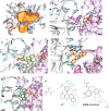
Fig 2. Molecular modeling studies of the acridinones series.
(A), steric complementarity between the colchicine site and S-7 (B), binding mode of S-11 showing a hydrogen bond between the NH groups of acridinones and Thr α179 (C), binding mode of S-10 where the trimethoxybenzyl group occupies the hydrophobic pocket of the colchicine site (D), docking pose for S-7 with a hydrogen bond between the NO2 group and Leu β252 (E), docking pose for S-6 with a hydrogen bond between oxygen from dioxalane and Cys β241 (F), Comparison of predicted interactions between the docking pose of S-7 and the crystal structure of tubulin (PDB ID: 1SA0) and the predicted interactions between crystallographic DAMA-colchicine (PDB ID: CN2) and tubulin (PDB ID: 1SA0). These interactions were calculated using the online software PoseView [38]. The green lines represent hydrophobic interactions and the black dotted lines represent hydrogen bonds.
Fig 3
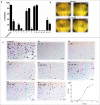
Fig 3. Effects of the acridinones on MDA-MB-231 cell migration.
(A), inhibition of wound closure (concentration of 10 μM) (B), pictures of the wound healing assay at 0 h and 22 h for the negative control and 10 (C), representative pictures of migrated cells in a Boyden chamber assay for the negative control and 10 in different concentrations (D), a representative IC50 curve for the antimigratory response against 10.
Fig 4
Fig 4. Comparison of cytotoxicity in MDA-MB-231 (black lines and circles) and FGH (red lines and circles) cells.
The results for 6 (A), 7 (B), 9 (C), 10 (D). The mean ± SD of two independent experiments in triplicate is shown, and the percentage of dead cells is relative to negative control.
Fig 5
Fig 5. In vitro tubulin assembly inhibition.
(A), results for the fluorescence-based assay; blue lines and circles represent the negative control (DMSO 1%), orange lines and squares represent the stabilizing control (paclitaxel) and green lines and triangles represent inhibition by the control (colchicine). 6 results are presented as purple lines and inverted triangles, 7 are dark red lines and hexagons and the results for 10 are presented as light red lines and circles (B), results for the light-scattering assay negative control, paclitaxel, colchicine, 6, 7 and 10 are represented by the same colors and shapes as in (A), 9 is presented as dark blue lines and hexagons (C), competitive colchicine binding assay; black lines and circles represent the fluorescence of the negative control (DMSO 1%) as competitor, and the results for positive control colchicine as competitor are presented as red lines and diamonds; fluorescence in the presence of 10 as competitor is presented as green lines and triangles. Data are representative of two experiments. *To monitor a non-competitive colchicine-binding behavior, experiments using an inactive compound from our in house collection of compounds were conducted (data not shown).
Fig 6
Fig 6. Flow cytometry analyses.
(A), the left panel presents the fluorescence histogram with the DNA profile of the cell-cycle phases (Sub-G1, G1, S and G2) of MDA-MB-231 cells in the presence of the control (DMSO 0.1%), colchicine and 10. The right panel shows the distribution of cells in different phases of the cell cycle for the control and after treatment with colchicine, 6, 7, 9 and 10 (B), the left panel displays the dot plot for the Annexin-V FITC/PI double stain of MDA-MB-231 cells for the control and after treatment with colchicine and 10. In the FITC- PI- quadrant are the alive cells, in the FITC+ quadrant are the apoptotic cells, and in the FITC+ PI+ and PI+ quadrants are the dead cells. The right panel shows the percentage of alive, apoptotic and dead cells of the control and after treatment with colchicine, 6, 7, 9 and 10. Statistical significance relative to control * P <0.05, ** P <0.01 and *** P <0.001.

References

    1. Wade RH. On and around microtubules: an overview. Mol Biotechnol. 2009;43: 177–91. 10.1007/s12033-009-9193-5 - DOI - PubMed
    1. Jordan MA, Wilson L. Microtubules as a target for anticancer drugs. Nat Rev Cancer. 2004;4: 253–65. - PubMed
    1. Checchi PM, Nettles JH, Zhou J, Snyder JP, Joshi HC. Microtubule-interacting drugs for cancer treatment. Trends Pharmacol Sci. 2003;24: 361–5. - PubMed
    1. Li Y-J, Qin Y-J, Makawana J a, Wang Y-T, Zhang Y-Q, Zhang Y-L, et al. Synthesis, biological evaluation and molecular modeling of 1,3,4-thiadiazol-2-amide derivatives as novel antitubulin agents. Bioorg Med Chem. 2014;22: 4312–22. 10.1016/j.bmc.2014.05.017 - DOI - PubMed
    1. Dumontet C, Jordan MA. Microtubule-binding agents: a dynamic field of cancer therapeutics. Nat Rev Drug Discov. 2010;9: 790–803. 10.1038/nrd3253 - DOI - PMC - PubMed

LinkOut - more resources