. 2016 Aug 9;24(2):246-55.
doi: 10.1016/j.cmet.2016.07.004.

Human and Mouse Brown Adipose Tissue Mitochondria Have Comparable UCP1 Function

Affiliations

Human and Mouse Brown Adipose Tissue Mitochondria Have Comparable UCP1 Function

Craig Porter et al. Cell Metab. .

Abstract

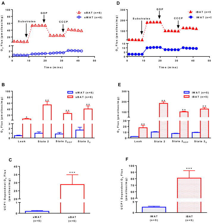
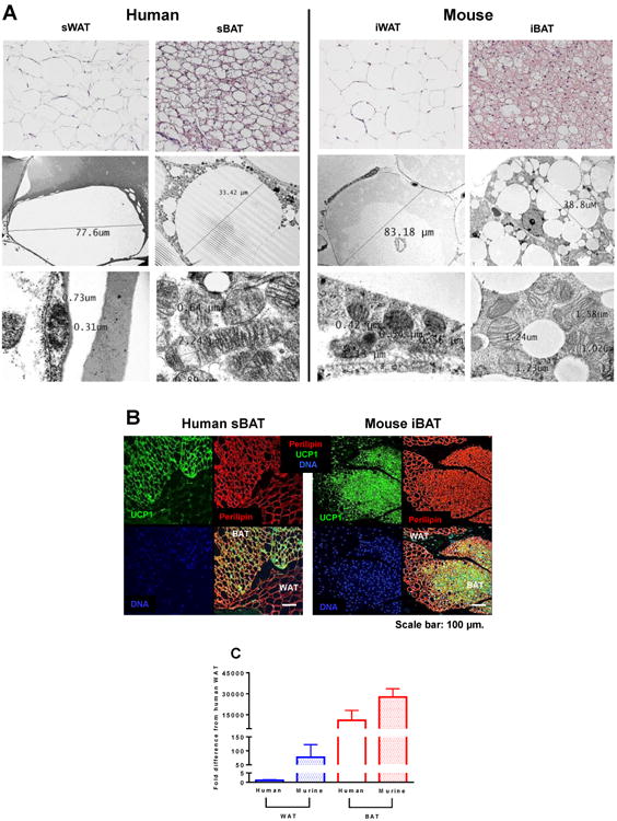
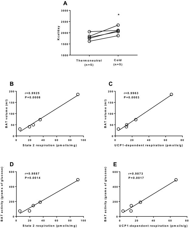
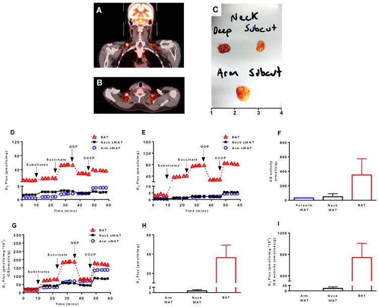
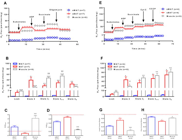
Brown adipose tissue (BAT) plays an important role in mammalian thermoregulation. The component of BAT mitochondria that permits this function is the inner membrane carrier protein uncoupling protein 1 (UCP1). To the best of our knowledge, no studies have directly quantified UCP1 function in human BAT. Further, whether human and rodent BAT have comparable thermogenic function remains unknown. We employed high-resolution respirometry to determine the respiratory capacity, coupling control, and, most importantly, UCP1 function of human supraclavicular BAT and rodent interscapular BAT. Human BAT was sensitive to the purine nucleotide GDP, providing the first direct evidence that human BAT mitochondria have thermogenically functional UCP1. Further, our data demonstrate that human and rodent BAT have similar UCP1 function per mitochondrion. These data indicate that human and rodent BAT are qualitatively similar in terms of UCP1 function.


Conflict of interest statement

The authors have no conflicts of interest to disclose.

Figures

Figure 1
Figure 1
Mitochondrial respirometry in human and rodent white and brown adipose tissue. (a) Representative respiration experiments performed on permeabilized sWAT and sBAT from humans following the sequential titration of substrates (1.5 mM octanoyl-l-carnitine, 5 mM pyruvate, 2 mM malate, 10 mM glutamate), 20 mM of the UCP1 inhibitor GDP and 5 μM of the protonophore CCCP (values are group means). (b) Comparison of respiratory states (state 2, State 2GDP, and State 2U) for human sWAT and sBAT shown in panel a. (c) UCP1-dependent respiration in human sWAT and sBAT calculated as the change in respiration following titration of the UCP1 inhibitor GDP. (d) Representative respiration experiments performed on permeabilized iWAT and iBAT from mice as described in panel a. (e) Comparison of respiratory states (state 2, State 2GDP, and State 2U) for mouse iWAT and iBAT shown in panel d. (f) UCP1-dependent respiration in mouse iWAT and iBAT calculated as in c. Note that leak respiration prior to titration of substrates is supported by endogenous substrates, likely FFAs. Values are presented as group means ± SEM unless otherwise stated. *P<0.05, **P<0.01, and **P<0.001 vs. WAT.
Figure 2
Figure 2
Morphological and molecular evaluation of human and rodent white and brown adipose tissue. (a) Representative hematoxylin and eosin stained sections of human sWAT and sBAT and rodent iWAT and iBAT (top row) showing differences in cell size and morphology between brown and white adipocytes form both humans and mice. Electron micrograph imaging humans WAT and sBAT and rodent iWAT and iBAT (middle row). Electron micrograph imaging of mitochondrial abundance and morphology of human sWAT and sBAT and rodent iWAT and iBAT (bottom row), showing the scarcity of mitochondrion in white adipose tissue in contrast to an abundance of large electron-dense organelles in brown adipose tissue. (b) Immunofluorescence staining of human sBAT and rodent iBAT for UCP1 (green), perilipin (red), and nuclei (blue) underscoring morphological differences between adipose tissue types and the presence of UCP1 in brown adipocytes. (c) UCP1 mRNA expression in human and rodent white and brown adipose tissue. Values are means ± SEM.
Figure 3
Figure 3
The metabolic significance of human brown adipose tissue. (a) Resting energy expenditure (REE) determined during thermoneutral conditions and mild non-shivering cold exposure in 5 healthy men. There was a significant (∼16%) increase in REE with mild non-shivering cold exposure (*P<0.05), likely the result of acute BAT activation. BAT volume (ml) determined by PET-CT significantly correlated with BAT mitochondrial respiratory capacity (b) and UCP1 function (c) in humans. BAT activity (total BAT glucose disposal) determined by PET-CT significantly correlated with BAT mitochondrial respiratory capacity (d) and UCP1 function (e) in humans. These data suggest that indices of BAT metabolic function in vivo are related to sBAT mitochondrial respiratory capacity and UCP1 function, although we acknowledge that the sample size is limit (n=5). It should be noted that there was also a strong correlation (r=0.998, P<0.001) between state 2 and UCP1-dependent respiration (data not shown), suggesting a relationship between BAT mitochondrial respiratory capacity and UCP1 function.
Figure 4
Figure 4
Functional evidence of symmetrical brown adipose tissue depots in humans. (a & b) PET-CT imaging showing that humans have symmetrical sBAT depots. (c) Sub-platysmal peri-jugular adipose tissue was sampled from both the left and right side of the neck of a severely burned individual. sWAT was sampled from the left and right sides of the neck above the platysma muscle, and forearm sWAT was also sampled from this patient. Panel c depicts the gross form and coloration of these three adipose tissue types, where deep neck fat depicts sub-platysmal peri-jugular adipose tissue and subcut neck depicts sWAT harvested above the platysma. (d) BAT and sWAT respirometry from the left side of the neck and sWAT from the forearm. (e) BAT and sWAT respirometry from the right side of the neck from the same patient shown in panel d (data from the same forearm sWAT sample shown in panel d is included for comparison). These data suggest that sBAT on either side of the neck contain mitochondria with functional UCP1. (f) Quantification of citrate synthase (CS) activity (a proxy of mitochondrial protein abundance) in sWAT from the forearm (n=1 sample), sWAT from the left and right side of the neck (n=2 samples), and BAT from both the left and right side of the neck (n=2 samples). (g) Mitochondrial respiration data presented in panels d and e normalized to CS activity data presented in panel f suggests that much (but not all) of the gradient in oxidative capacity between human WAT and BAT may be explained by mitochondrial protein abundance. (h) Mass specific UCP1-dependent respiration (respiration per mg of tissue) in neck BAT and sWAT and forearm sWAT. (i) Mitochondria specific UCP1-dependent respiration (respiration per unit of CS activity) in neck BAT and sWAT and forearm sWAT. These results suggest that whether presented per unit of tissue or mitochondrial protein, UCP1 function is only apparent in sBAT of humans. Note that leak respiration prior to titration of substrates is supported by endogenous substrates, likely FFAs.
Figure 5
Figure 5
Comparison of mitochondrial respiratory capacity and coupling control in human sWAT, sBAT, and skeletal muscle. (a) Representative respiration experiments performed on permeabilized sWAT and sBAT from the same seven individuals and skeletal muscle from six separate individuals following the sequential titration of substrates and oligomycin as describe in Fig 1A (values are group means). (b) Comparison of respiratory states for sWAT, sBAT and skeletal muscle shown in panel a highlight similar respiratory capacity in human BAT and skeletal muscle. (c) The respiratory control ratio for ADP (RCRADP) calculated by dividing state 3I by state 2. A RCRADP < 1 indicates that mitochondria are not coupled in human sBAT. (d) The coupling control ratio for oligomycin (CCROM) calculated by dividing state 4O by state 3I+II. A CCROM of = 1 indicates that mitochondria are insensitive to oligomycin and thus are uncoupled. A lower CCROM suggest better coupled mitochondria in sWAT and skeletal muscle. (e) Representative respiration experiments performed on permeabilized sWAT, sBAT and skeletal muscle from the same individuals in panel a following the sequential titration of substrates (1.5 mM octanoyl-l-carnitine, 5 mM pyruvate, 2 mM malate, and 10 mM glutamate), ADP (5 mM), succinate (10 mM), cytochrome C (10 μM) and the ionophore CCCP (5 μM) (values are group means). (f) Comparison of respiratory states for sWAT, sBAT and skeletal muscle shown in panel e again showing that sBAT has a respiratory capacity more akin to that of muscle than sWAT in humans. (g) The leak control ratio (LCR), calculated by dividing state 3I+II by state 3U, shows the absence of leak control in human sBAT. The lux control ratio (FCR), an index of the efficiency of the oxidative phosphorylation system, was calculated by dividing state 2 by state 3U. A lower FCR in BAT demonstrates poor mitochondrial coupling control. Note that while the addition of ADP to sBAT reduces leak respiration there is also likely a stimulation of coupled respiration (ATP production). However, this is minimal given the blunted response to the ATP synthase inhibitor oligomycin observed in coupled sBAT mitochondria. Note that leak respiration prior to titration of substrates is supported by endogenous substrates, likely FFAs. Values are presented as means ± SE unless otherwise stated. *P<0.05 vs. sWAT; **P<0.01 vs. sWAT; ***P<0.001 vs. sWAT; P<0.05 vs. sBAT; ††P<0.01 vs. sBAT; †††P<0.001 vs. sBAT.

References

    1. Chondronikola M, Annamalai P, Chao T, Porter C, Saraf M, Cesani F, Sidossis L. A percutaneous needle biopsy technique for sampling the supraclavicular brown adipose tissue depot of humans. Int J Obes (Lond) 2015 Epub ahead of print. - PMC - PubMed
    1. Chondronikola M, Volpi E, Børsheim E, Chao T, Porter C, Annamalai P, Yfanti C, Labbe S, Hurren N, Malagaris I, et al. Brown Adipose Tissue Is Linked to a Distinct Thermoregulatory Response to Mild Cold in People. Front Physiol. 2016a;19:7, 129. - PMC - PubMed
    1. Chondronikola M, Volpi E, Børsheim E, Porter C, Annamalai P, Enerbäk S, Liddell ME, Saraf M, Labbe S, Hurren S, et al. Brown adipose tissue activation improves glucose homeostasis and insulin sensitivity in humans. Diabetes. 2014;63:4089–4099. - PMC - PubMed
    1. Chondronikola M, Volpi E, Børsheim E, Porter C, Saraf M, Annamalai P, Yfanti C, Chao T, Wong D, Shinoda K, et al. Brown Adipose Tissue Activation Is Linked to Distinct Systemic Effects on Lipid Metabolism in Humans. Cell Metab. 2016b;23(6):1200–1206. - PMC - PubMed
    1. Cypess A, White A, Vernochet C, Schulz T, Xue R, Sass C, Huang T, Roberts-Toler C, Weiner L, Sze C, et al. Anatomical localization, gene expression profiling and functional characterization of adult human neck brown fat. Nat Med. 2013a;19 - PMC - PubMed

Publication types

Substances