. 2016 Nov;25(11):1977-1988.
doi: 10.1002/pro.3010. Epub 2016 Aug 23.

Intrinsic protein disorder could be overlooked in cocrystallization conditions: An SRCD case study

Affiliations

Intrinsic protein disorder could be overlooked in cocrystallization conditions: An SRCD case study

Eszter Németh et al. Protein Sci. 2016 Nov.

Abstract

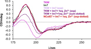
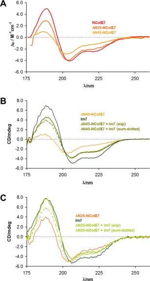
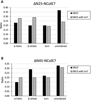
X-ray diffractometry dominates protein studies, as it can provide 3D structures of these diverse macromolecules or their molecular complexes with interacting partners: substrates, inhibitors, and/or cofactors. Here, we show that under cocrystallization conditions the results could reflect induced protein folds instead of the (partially) disordered original structures. The analysis of synchrotron radiation circular dichroism spectra revealed that the Im7 immunity protein stabilizes the native-like solution structure of unfolded NColE7 nuclease mutants via complex formation. This is consistent with the fact that among the several available crystal structures with its inhibitor or substrate, all NColE7 structures are virtually the same. Our results draw attention to the possible structural consequence of protein modifications, which is often hidden by compensational effects of intermolecular interactions. The growing evidence on the importance of protein intrinsic disorder thus, demands more extensive complementary experiments in solution phase with the unligated form of the protein of interest.

Keywords: Im7 immunity protein; SRCD spectroscopy; colicin E7 nuclease; induced protein folding.


Figures

Figure 1
Figure 1
Scheme of the NColE7 variant sequences: the sequences of the purified NColE7 and Im7 proteins are also shown. The GPLGSPEF is an additional sequence encoded by the pGEX‐6P‐1 plasmid, which remained at the N‐terminus of the nucleases after enzymatic cleavage of the GST purification tag. TKW symbolizes the TKW, TK, KW, or W NColE7 point mutants, in which T454A and/or K458A and/or W464A mutations are present. For the truncated mutants X in the ΔNX notation indicates the number of deleted amino acids at their N‐termini. The Im7 protein is purified together with a hexahistidine tag at its C‐terminus.
Figure 2
Figure 2
Consequences of NColE7–Im7 interaction. (A) The comparison of the structures of free and bound Im7 and NColE7 proteins visualized from their single crystal structures. The free NColE7 structure (green, 1M08 15), as well as the free Im7 structure (yellow, 1CEI 27) is superimposed to the structure of the NColE7‐Im7 complex (blue, 7CEI 28). The Zn2+‐ions are indicated by spheres. (B) The experimental SRCD spectra of NColE7 and Im7 proteins (3.2 × 10⁻⁵ M) and their equimolar mixtures (1.6 × 10⁻⁵ M, each). The mathematical sum of the component protein spectra considering the same composition as in the experiment, but assuming no interaction between the two proteins is also shown for comparison. (C) The same as (B) but the spectra were recorded in the presence of an equivalent amount of Zn2+‐ions referred to the actual NColE7 concentration.
Figure 3
Figure 3
Im7 binding of truncated NColE7 mutants. The result of the GST pull‐down assay showing the interaction of Im7 with N‐terminally truncated NColE7 mutants in their GST‐tagged forms. The first lane contains the Thermo Scientific™ Unstained Protein Molecular Weight Marker. In lanes 3–5 the NColE7 proteins were shown to bind Im7 under the conditions of the GST‐affinity purification, while the shortest mutant protein in lane 2 did not.
Figure 4
Figure 4
DNA binding of ΔN45‐NColE7 mutant. Flow linear dichroism spectra of a 130 μM (referred to base pairs) CT‐DNA sample containing Zn2+‐ions (50 µM) in the presence and absence of ΔN45‐NColE7 (5 µM). The spectra are compared with those of the same DNA sample incubated with increasing amounts of NColE7 (0.5–1.5 µM) in the presence of 60 µM EDTA to avoid DNA cleavage by the active enzyme.
Figure 5
Figure 5
Induction of the native‐like structure of the TKW mutant in solution. Comparison of the experimental SRCD spectra of TKW mutant, NColE7, and Im7 proteins (3.2 × 10⁻⁵ M) and the equimolar mixtures of the nucleases and Im7 (1.6 × 10⁻⁵ M, each). The calculated spectral sum was constructed from the component spectra according to the initial composition of the measured system—thus, representing a spectrum of a mixture without interaction between the TKW mutant and Im7.
Figure 6
Figure 6
SRCD spectra of the N‐terminally truncated NColE7 mutants. (A) Comparison of the molar SRCD spectra of the ΔN25, ΔN45‐NColE7 truncated mutant proteins and that of the native NColE7. The calculation of the Δε values was based on the average amino acid residue molecular weight for each protein. (B) Experimental spectra of Im7 protein and the ΔN45‐NColE7 mutant (3.2 × 10⁻⁵ M, each) and their equimolar mixtures (1.6 × 10⁻⁵ M, each). The calculated spectra constructed from the component spectra are also shown for comparison. (C) Comparison of the experimental spectra of Im7 and the ΔN25‐NColE7 mutant proteins (3.2 × 10⁻⁵ M, each) and their equimolar mixtures (1.6 × 10⁻⁵ M, each). The calculated spectra constructed from the component spectra are also shown.
Figure 7
Figure 7
The comparison of the induced folding in the ΔNX‐NColE7–Im7 systems: change in the distribution of secondary structure elements of ΔNX‐NColE7 mutants as a consequence of the interaction with Im7 protein as calculated by the CDPro program package. The data on the NColE7 variants in complex with Im7 were obtained from the difference spectra of the complex and Im7 with the asumption that the structure of Im7 is stable and does not significantly change on complex formation. (A) ΔN25‐NColE7 mutant; (B) ΔN45‐NColE7 mutant.

References

    1. Németh E, Körtvélyesi T, Kožíšek M, Thulstrup PW, Christensen HEM, Asaka MN, Nagata K, Gyurcsik B (2014) Substrate binding activates the designed triple mutant of the colicin E7 metallonuclease. J Biol Inorg Chem 19:1295–1303. - PubMed
    1. Németh E, Kožíšek M, Schilli GK, Gyurcsik B (2015) Preorganization of the catalytic Zn2+‐binding site in the HNH nuclease motif—a solution study. J Inorg Biochem 151:143–149. - PubMed
    1. Walker D, Lancaster L, James R, Kleanthous C (2004) Identification of the catalytic motif of the microbial ribosome inactivating cytotoxin colicin E3. Protein Sci 13:1603–1611. - PMC - PubMed
    1. Papadakos G, Sharma A, Lancaster LE, Bowen R, Kaminska R, Leech AP, Walker D, Redfield C, Kleanthous C (2015) Consequences of inducing intrinsic disorder in a high‐affinity protein−protein interaction. J Am Chem Soc 137:5252–5255. - PubMed
    1. Chak KF, Kuo WS, Lu FM, James R (1991) Cloning and characterization of the ColE7 plasmid. J Gen Microbiol 137:91–100. - PubMed

Publication types

Substances

LinkOut - more resources