. 2016 Jun 21;7(25):38427-39.
doi: 10.18632/oncotarget.9538.

Loss of GFAT1 promotes epithelial-to-mesenchymal transition and predicts unfavorable prognosis in gastric cancer

Affiliations

Loss of GFAT1 promotes epithelial-to-mesenchymal transition and predicts unfavorable prognosis in gastric cancer

Fangfang Duan et al. Oncotarget. .

Abstract

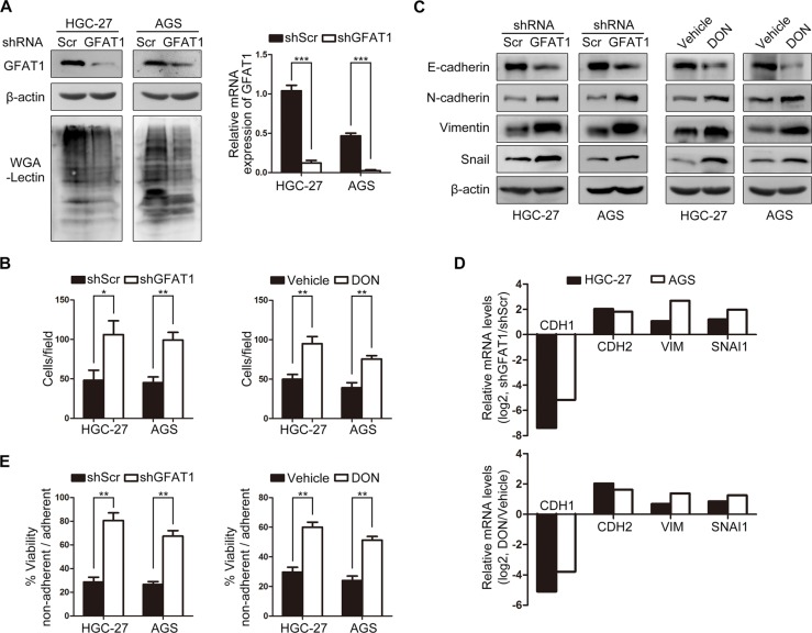
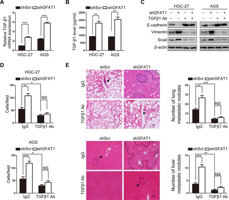
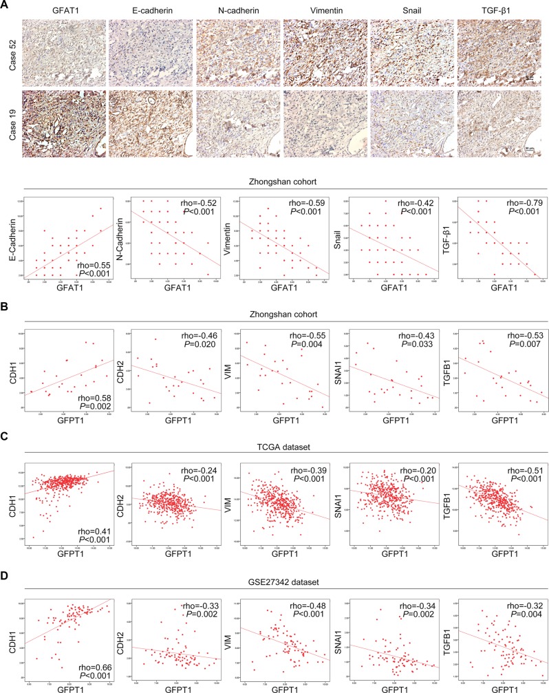
Gastric cancer remains the third leading cause of cancer-related mortality worldwide, and invasion and metastasis of gastric cancer represent the major reason for its poor prognosis. Glutamine: fructose-6-phosphate amidotransferase 1 (GFAT1) is the first and rate-limiting enzyme of hexosamine biosynthesis pathway (HBP). Nevertheless, the role of GFAT1 in gastric cancer is little investigated. In this study, we found that the expression of GFAT1 was decreased in gastric cancer. Low expression of GFAT1 was positively associated with vessel invasion, late T stage, lymph node metastasis, distant metastasis, advanced TNM stage and poor prognosis in patients with gastric cancer. Furthermore, in vitro and in vivo studies revealed that down-regulation of GFAT1 promoted epithelial-to-mesenchymal transition (EMT) and invasive activities in gastric cancer cells through inducing the expression of TGF-β1. The GFAT1 expression also significantly correlated with EMT-related factors in gastric cancer patients. Together, these findings indicate that GFAT1 functions as a novel suppressor of EMT and tumor metastasis in gastric cancer.

Keywords: GFAT1; TGF-β1; epithelial-to-mesenchymal transition; gastric cancer; prognostic factor.


Conflict of interest statement

The authors declare no competing interests.

Figures

Figure 1
Figure 1. The expression of GFAT1 is decreased in gastric cancer
(A) The mRNA expression of GFPT1 in 25 pairs of gastric cancer tissues and adjacent normal mucosa was examined by real-time PCR analysis. Data shown are the log values of tumor vs normal. (B) Expression profiling of GFPT1 mRNA in matched gastric cancer samples from GSE27342 and GSE13911 datasets. Data shown are the log values of tumor vs normal. (C) GFAT1 protein expression and WGA lectin staining in 25 paired gastric cancer samples was detected by western-blot. Blots shown are representative results in 6 cases. N, adjacent non-tumor sections; T, tumor sections. (D) GFAT1 protein expression in 15 paired gastric cancer samples was detected by IHC. Images shown are representative of GFAT1 staining in 2 paired gastric cancer tissues. The expression of GFAT1 in paired tumor tissues was compared by IHC scoring. In (B–C), data are represented as Means ± SD. In (D), data are represented as min-to-max bar graphs with median lines.
Figure 2
Figure 2. Kaplan-Meier survival analysis for overall survival of gastric cancer patients according to the GFAT1 expression
(AB) The association of GFAT1 expression with overall survival was examined by Kaplan-Meier analysis in gastric cancer patients from Zhongshan cohort (A) and TCGA dataset (B), respectively. Total patients in each cohort were also divided into TNM I-II stage and TNM III-IV stage subgroups for analysis. (C) The association of GFAT1 expression with overall survival and progression-free survival was examined by Kaplan-Meier analysis in gastric cancer patients using the online survival analysis software (http://www.kmplot.com/analysis/index.php?p=service&cancer=gastric).
Figure 3
Figure 3. GFAT1 suppresses epithelial-to-mesenchymal transition and invasive activities in gastric cancer cells
(A) The GFAT1 expression pattern and WGA lectin staining in gastric cancer cells stably transfected with scramble (Scr) or GFAT1 shRNA. (B) The effect of GFAT1 specific shRNA or GFAT1 inhibitor DON on the invasion of gastric cancer HGC-27 and AGS cells by transwell analysis. (CD) The effect of GFAT1 specific shRNA or GFAT1 inhibitor DON on the expression of EMT markers in gastric cancer HGC-27 and AGS cells was evaluated by western-blot (C) and real-time PCR (D) analysis, respectively. (E) The effect of GFAT1 specific shRNA or GFAT1 inhibitor DON on the relative viability of gastric cancer HGC-27 and AGS cells by anoikis assay. *P < 0.05; **P < 0.01; ***P < 0.001. In (A–B) and (E), data are represented as Means ± SD.
Figure 4
Figure 4. Loss of GFAT1 promotes epithelial-to-mesenchymal transition through inducing TGF-β1 expression in gastric cancer
(A) The effect of GFAT1 specific shRNA on the mRNA expression of TGF-β1 in gastric cancer HGC-27 and AGS cells was detected by real-time PCR analysis. (B) The effect of GFAT1 specific shRNA on the levels of TGF-β1 in the culture supernatant of gastric cancer HGC-27 and AGS cells was detected by ELISA. (C) The effect of TGF-β1 neutralizing antibody on the expression of EMT markers in control or GFAT1-depleted gastric cancer cells was examined by western blot. (D) The effects of TGF-β1 neutralizing antibody on the invasion of control or GFAT1-depleted HGC-27 and AGS cells were examined by transwell analysis. (E) Stable AGS cells were injected into the lateral tail vein of mice. After the sacrifice of mice, lung and liver tissues was collected. Images were representatives of HE staining in lung (upper panel) and liver (lower panel) tissue sections from each group (scale bar, 50 μm). Micrometastatic lesions were indicated with arrows or dotted line. Numbers of lung and liver metastatic foci in each group were also counted. *P < 0.05; **P < 0.01; ***P < 0.001; NS, no significance. In (A–B) and (D–E), data are represented as Means ± SD.
Figure 5
Figure 5. Correlated expression of GFAT1 with EMT-related factors in gastric cancer
(A) The expression of EMT-related factors E-cadherin, N-cadherin, Vimentin, Snail and TGF-β1 in gastric cancer tissues from Zhongshan cohort was examined by IHC, and the correlation between their expression and GFAT1 was assessed individually. Representative images of IHC staining were shown in the upper panel. (BD) The correlated expression of GFPT1 mRNA level with transcript levels of CDH1, CDH2, VIM, SNAI1 and TGFB1 in Zhongshan cohort (B) as well as in TCGA (C) and GSE27342 (D) datasets.

References

    1. Siegel R, Ma J, Zou Z, Jemal A. Cancer statistics, 2014. CA Cancer J Clin. 2014;64:9–29. - PubMed
    1. Torre LA, Bray F, Siegel RL, Ferlay J, Lortet-Tieulent J, Jemal A. Global cancer statistics, 2012. CA Cancer J Clin. 2015;65:87–108. - PubMed
    1. Ferlay J, Shin HR, Bray F, Forman D, Mathers C, Parkin DM. Estimates of worldwide burden of cancer in 2008: GLOBOCAN 2008. Int J Cancer. 2010;127:2893–2917. - PubMed
    1. Ferlay J, Steliarova-Foucher E, Lortet-Tieulent J, Rosso S, Coebergh JW, Comber H, Forman D, Bray F. Cancer incidence and mortality patterns in Europe: estimates for 40 countries in 2012. Eur J Cancer. 2013;49:1374–1403. - PubMed
    1. Washington K. 7th edition of the AJCC cancer staging manual: stomach. Ann Surg Oncol. 2010;17:3077–3079. - PubMed

MeSH terms

Substances