. 2016 Sep 23;291(39):20729-38.
doi: 10.1074/jbc.M116.752485. Epub 2016 Aug 10.

Phosphatidic Acid Produced by RalA-activated PLD2 Stimulates Caveolae-mediated Endocytosis and Trafficking in Endothelial Cells

Affiliations

Phosphatidic Acid Produced by RalA-activated PLD2 Stimulates Caveolae-mediated Endocytosis and Trafficking in Endothelial Cells

Ying Jiang et al. J Biol Chem. .

Abstract

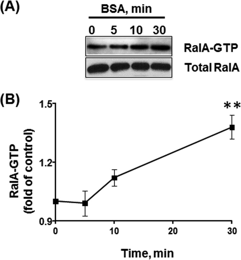
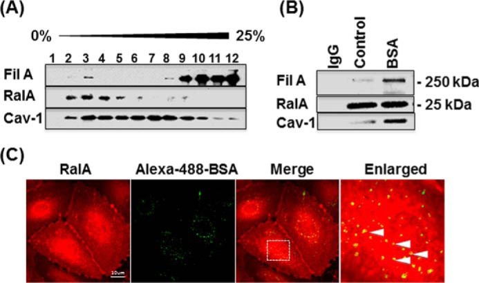
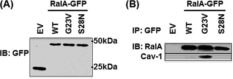
Caveolae are the primary route for internalization and transendothelial transport of macromolecules, such as insulin and albumin. Caveolae-mediated endocytosis is activated by Src-dependent caveolin-1 (Cav-1) phosphorylation and subsequent recruitment of dynamin-2 and filamin A (FilA), which facilitate vesicle fission and trafficking, respectively. Here, we tested the role of RalA and phospholipase D (PLD) signaling in the regulation of caveolae-mediated endocytosis and trafficking. The addition of albumin to human lung microvascular endothelial cells induced the activation of RalA within minutes, and siRNA-mediated down-regulation of RalA abolished fluorescent BSA uptake. Co-immunoprecipitation studies revealed that albumin induced the association between RalA, Cav-1, and FilA; however, RalA knockdown with siRNA did not affect FilA recruitment to Cav-1, suggesting that RalA was not required for FilA and Cav-1 complex formation. Rather, RalA probably facilitates caveolae-mediated endocytosis by activating downstream effectors. PLD2 was shown to be activated by RalA, and inhibition of PLD2 abolished Alexa-488-BSA uptake, indicating that phosphatidic acid (PA) generated by PLD2 may facilitate caveolae-mediated endocytosis. Furthermore, using a PA biosensor, GFP-PASS, we observed that BSA induced an increase in PA co-localization with Cav-1-RFP, which could be blocked by a dominant negative PLD2 mutant. Total internal reflection fluorescence microscopy studies of Cav-1-RFP also showed that fusion of caveolae with the basal plasma membrane was dependent on PLD2 activity. Thus, our results suggest that the small GTPase RalA plays an important role in promoting invagination and trafficking of caveolae, not by potentiating the association between Cav-1 and FilA but by stimulating PLD2-mediated generation of phosphatidic acid.

Keywords: Ras protein; caveolin; endocytosis; filamin; trafficking.


Figures

FIGURE 1.
FIGURE 1.
RalA activation induced by BSA. A, serum-deprived HLMVECs were stimulated with 30 mg/ml BSA for 5, 10, and 30 min, and RalA activity was assessed by RalBP1 pull-down assay. The amount of active GTP-bound RalA co-precipitated with the RalA-binding domain of RalBP1 was analyzed by Western blotting. Note the increase in RalA activity following 30 min of BSA stimulation. B, graph represents the time course of RalA activation induced by BSA, and the data are expressed as a percentage of the RalA activity in unstimulated (control) cells (mean ± S.E., n = 6). BSA stimulation induced a 35% increase in RalA activity.
FIGURE 2.
FIGURE 2.
RalA is associated with caveolin-1 and filamin A during caveolae-mediated endocytosis. A, analysis of RalA distribution in sucrose density gradient fractions of HLMVECs revealed the majority of RalA in fractions 2–4 associated with buoyant fractions (fractions 2–4) that also contain Cav-1 and FilA. B, serum-deprived HLMVECs were stimulated with vehicle alone or 30 mg/ml BSA for 30 min, lysed, clarified, immunoprecipitated with anti-RalA antibody versus control IgG, and blotted for Cav-1 and FilA. Cav-1 and FilA interaction with RalA was dramatically increased upon stimulation with BSA, suggesting formation of the protein complex during caveolae-mediated endocytosis. C, serum-deprived HLMVECs were incubated with Alexa-488-BSA for 30 min, acid-washed, fixed, and stained for RalA. Co-localization of endogenous RalA with internalized BSA (see enlarged merged image) implies that RalA remains associated with internalized, BSA-loaded caveolae. Images are representative of three independent experiments.
FIGURE 3.
FIGURE 3.
Nucleotide-dependent interaction between RalA and caveolin-1. HLMVECs transfected with EV or GFP-tagged RalA constructs were lysed 48 h after transfection and blotted with anti-GFP antibody. A, expression level of WT, constitutively active (G23V), and dominant negative (S28N) GFP-tagged RalA analyzed by Western blotting (IB) shows equal expression. B, GFP-tagged proteins were immunoprecipitated (IP) with anti-GFP antibody and blotted for RalA and caveolin-1. Note that Cav-1 associated with GTP-bound constitutively active RalA mutant. Results are representative of two independent experiments.
FIGURE 4.
FIGURE 4.
Effect of RalA mutants on caveolae-mediated endocytosis. A, HLMVECs were transfected with EV, GFP-tagged RalA, constitutively active RalA (G23V), or dominant negative RalA (S28N) mutants. Forty-eight h after transfection, cells were starved, incubated with 0.1 mg/ml BSA + 50 μg/ml Alexa-555-BSA for 30 min, acid-washed, and fixed, and confocal images of GFP and Alexa-555 BSA fluorescence were collected. Alexa-555-BSA images of transfected and non-transfected (NTF) cells were analyzed using ImageJ software (note the image mask of transfected cells). B, quantification of Alexa-555-BSA uptake. Dominant negative RalA (S28N) mutant reduced BSA endocytosis by 35% in comparison with non-transfected cells and cells expressing EV, WT RalA, or constitutively active (G23V) RalA. Data are mean ± S.E. (error bars) (n = 5); **, p < 0.01 versus EV by ANOVA.
FIGURE 5.
FIGURE 5.
Effect of filamin A siRNA on RalA activation and interaction with caveolin-1 and effect of RalA siRNA on interaction of filamin A and caveolin-1. A, HLMVECs transfected with control or FilA siRNA were stimulated with 30 mg/ml BSA for 30 min and lysed. Lysates were incubated with the RalA binding domain of RalBP1, and the amount of RalA-GTP was estimated by Western blotting. Quantification of RalA-GTP is shown in the graph (n = 3/group; *, p < 0.05 versus control siRNA + BSA 0 min; #, p < 0.05 versus FilA siRNA + BSA 0 min by ANOVA). B, starved or BSA-stimulated cells transfected with control or FilA siRNA were lysed, and RalA was immunoprecipitated from the whole cell lysates. Quantification of co-immunoprecipitated Cav-1 is shown in the graph (n = 3/group; *, p < 0.05 versus control siRNA + BSA 0 min by ANOVA). C, Western blotting analysis of RalA and Cav-1 in HLMVECs treated 48 h with SMART POOL (SP) RalA siRNA or each of four oligonucleotides separately versus scrambled control siRNA (Cont). D, HLMVECs transfected with control or RalA siRNA were stimulated with 30 mg/ml BSA for 30 min and lysed, and then FilA was immunoprecipitated. Immunoblotting for caveolin-1 revealed that knocking down RalA did not have a significant effect on Cav-1/FilA association. Results shown in C and D are representative of three independent experiments. IP, immunoprecipitation; IB, immunoblotting. AU, arbitrary units.
FIGURE 6.
FIGURE 6.
RalA siRNA and PLD inhibitors block caveolae-mediated endocytosis of BSA. A, starved HLMVECs treated with SMART POOL (SP) RalA siRNA or individually with each of the four oligonucleotides for 48 h were incubated with Alexa-488-BSA for 30 min, acid-washed, and fixed. Images of internalized fluorescent albumin were acquired by confocal microscopy. B, quantification of fluorescence intensity of Alexa-488-BSA demonstrates that RalA siRNA blocked BSA uptake (n = 10/group; ***, p < 0.001 versus siCont by ANOVA). C, HLMVECs treated with SMART POOL RalA siRNA for 48 h were transfected with RalA-GFP to rescue RalA expression as compared with GFP, which was transfected as a negative control. Cells were then lysed and examined by Western blotting (IB) to confirm RalA rescue. D, quantification of fluorescence intensity of Alexa-555-BSA uptake in RalA-depleted and -repleted HLMVECs (n = 10/group; ***, p < 0.001 by ANOVA). E, quantification of fluorescence intensity of Alexa-488-BSA demonstrates that both RalA knockdown by siRNA and 1-butanol dramatically inhibited BSA uptake (n = 6/group; ***, p < 0.001 versus control siRNA by ANOVA). F, HLMVECs treated with 200 nm VU0359595 (PLD1 inhibitor) or VU0364739 (PLD2 inhibitor) for 1 h were incubated with Alexa-488-BSA for 30 min. Quantification of Alexa-488-BSA fluorescence indicates that PLD2 inhibitor but not PLD1 inhibitor blocked albumin uptake (n = 7/group; ***, p < 0.001 versus control by ANOVA). Error bars, S.E.
FIGURE 7.
FIGURE 7.
Cav-1-RFP-positive vesicle trafficking and fusion. A, Western blotting analysis of overexpressed hPLD1 K898R (PLD1 mutant) and mPLD2 K758R (PLD2 mutant) versus EV and control cells. B, HLMVECs were infected with EV, hPLD1 K898R, or mPLD2 K758R mutant in adenovirus, labeled with [32P]ATP, and stimulated with thrombin (0.005 units/ml). [32P]PBt formation as a result of PLD activation indicated that PLD2 mutant inhibited thrombin-induced PLD activity, whereas PLD1 mutant had no effect. n = 3/group; *, p < 0.05; **, p < 0.01 by ANOVA. C, HLMVECs transfected with Cav-1-RFP and infected with EV, hPLD1-K898R, or mPLD2 K758R mutant were serum-deprived for 2 h, treated with 30 mg/ml BSA, and then imaged by live cell TIRF microscopy every 5 min for 20 min. Note the time-dependent appearance of Cav-1-RFP in the TIRF plane (abluminal aspect of the cell). D, quantification of relative fluorescence intensity of Cav-1-RFP (n = 3 regions/group from three independent experiments; **, p < 0.01 versus control; ***, p < 0.001 versus EV control + BSA by ANOVA). E, HLMVECs transfected with Cav-1-RFP and after 24 h were serum-deprived for 2 h, treated with 200 nm VU0359595 (PLD1 inhibitor) or VU0364739 (PLD2 inhibitor) for 1 h, and then stimulated with 30 mg/ml BSA and imaged by live cell TIRF microscopy every 5 min for 20 min (n = 3 regions/group from three independent experiments; ***, p < 0.001 versus control without BSA; **, p < 0.01 versus control with BSA by ANOVA). Error bars, S.E. AU, arbitrary units.
FIGURE 8.
FIGURE 8.
Effect of PLD2 mutant on PA generation in caveolae. A, HLMVECs were co-transfected with GFP-PASS and Cav-1-RFP following infection with EV, hPLD1-K898R, or mPLD2 K758R mutant; serum-deprived for 2 h; treated with 30 mg/ml BSA for 30 min; and imaged by confocal microscopy. B, confocal images representative of three independent experiments were used to determine Pearson's correlation coefficient of co-localized GFP-PASS and Cav-1-RFP fluorescence. Results indicate that BSA increases and PLD2 mutant reduces co-localization of PA and Cav-1 (n = 10/group; ***, p < 0.001 versus EV + BSA by ANOVA). Error bars, S.E.
FIGURE 9.
FIGURE 9.
Proposed model for role of RalA in caveolae-mediated endocytosis. In the presence of albumin, RalA is recruited to caveolae, where it associates with Cav-1 and FilA, ultimately becoming activated upon GTP binding. Activated RalA triggers downstream effector PLD2-mediated generation of PA, which promotes caveolae-mediated endocytosis and transcytosis.

Similar articles

Cited by

References

    1. Parton R. G., and del Pozo M. A. (2013) Caveolae as plasma membrane sensors, protectors and organizers. Nat. Rev. Mol. Cell Biol. 14, 98–112 - PubMed
    1. Ahn S., Maudsley S., Luttrell L. M., Lefkowitz R. J., and Daaka Y. (1999) Src-mediated tyrosine phosphorylation of dynamin is required for β2-adrenergic receptor internalization and mitogen-activated protein kinase signaling. J. Biol. Chem. 274, 1185–1188 - PubMed
    1. Ahn S., Kim J., Lucaveche C. L., Reedy M. C., Luttrell L. M., Lefkowitz R. J., and Daaka Y. (2002) Src-dependent tyrosine phosphorylation regulates dynamin self-assembly and ligand-induced endocytosis of the epidermal growth factor receptor. J. Biol. Chem. 277, 26642–26651 - PubMed
    1. Shajahan A. N., Timblin B. K., Sandoval R., Tiruppathi C., Malik A. B., and Minshall R. D. (2004) Role of Src-induced dynamin-2 phosphorylation in caveolae-mediated endocytosis in endothelial cells. J. Biol. Chem. 279, 20392–20400 - PubMed
    1. Zimnicka A. M., Husain Y. S., Shajahan A. N., Sverdlov M., Chaga O., Chen Z., Toth P. T., Klomp J., Karginov A. V., Tiruppathi C., Malik A. B., and Minshall R. D. (2016) Src-dependent phosphorylation of caveolin-1 Tyr14 promotes swelling and release of caveolae. Mol. Biol. Cell 27, 2090–2106 - PMC - PubMed

Publication types

MeSH terms

LinkOut - more resources