Review
. 2017 Mar:171:93-103.
doi: 10.1016/j.pharmthera.2016.08.003. Epub 2016 Aug 7.

Pericytes for the treatment of orthopedic conditions

Affiliations
Review

Pericytes for the treatment of orthopedic conditions

Aaron W James et al. Pharmacol Ther. 2017 Mar.

Abstract

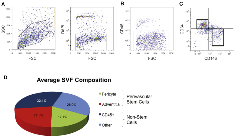
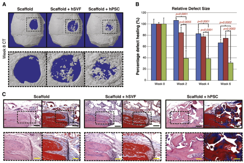
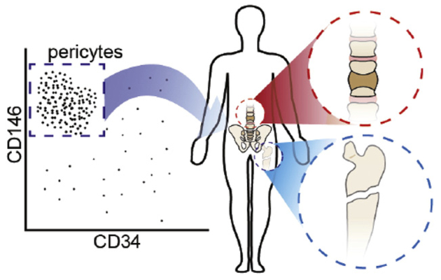
Pericytes and other perivascular stem cells are of growing interest in orthopedics and tissue engineering. Long regarded as simple regulators of angiogenesis and blood pressure, pericytes are now recognized to have MSC (mesenchymal stem cell) characteristics, including multipotentiality, self-renewal, immunoregulatory functions, and diverse roles in tissue repair. Pericytes are typified by characteristic cell surface marker expression (including αSMA, CD146, PDGFRβ, NG2, RGS5, among others). Although alone no marker is absolutely specific for pericytes, collectively these markers appear to selectively identify an MSC-like pericyte. The purification of pericytes is most well described as a CD146+CD34-CD45- cell population. Pericytes and other perivascular stem cell populations have been applied in diverse orthopedic applications, including both ectopic and orthotopic models. Application of purified cells has sped calvarial repair, induced spine fusion, and prevented fibrous non-union in rodent models. Pericytes induce these effects via both direct and indirect mechanisms. In terms of their paracrine effects, pericytes are known to produce and secrete high levels of a number of growth and differentiation factors both in vitro and after transplantation. The following review will cover existing studies to date regarding pericyte application for bone and cartilage engineering. In addition, further questions in the field will be pondered, including the phenotypic and functional overlap between pericytes and culture-derived MSC, and the concept of pericytes as efficient producers of differentiation factors to speed tissue repair.

Keywords: Bone; Cartilage; MSC; Mesenchymal stem cell; PSC; Perivascular stem cell.


Conflict of interest statement

Conflict of interest statement

K.T. and C.S. are inventors of perivascular stem cell-related patents filed from UCLA. K.T. and C.S. are founders of Scarless Laboratories, Inc. which sublicenses perivascular stem cell-related patents from the UC Regents, and who also hold equity in the company. C.S. is also an officer of Scarless Laboratories, Inc.

Figures

Fig. 1.
Fig. 1.
Schematic overview of past studies involving orthopedic applications of pericyte and perivascular stem cells (PSC). From left to right: Pericytes and perivascular stem cells are most commonly derived from liposuction aspirate of human white adipose tissue. After enzymatic digestion, an unpurified stromal cell population is obtained, termed ‘stromal vascular fraction’ or SVF. Fluorescence activated cell sorting is performed to obtain CD146+CD34CD45 pericytes and CD146CD34+CD45 adventitial cells. Collectively, these perivascular cell populations are termed ‘perivascular stem cell’ or PSC. Pericytes and/or PSC have been applied to diverse orthopedic models, including mouse calvarial defects, mouse intramuscular implantation experiments, rat femoral fibrous non-union defects, and rat lumbar transverse spinal fusion models.
Fig. 2.
Fig. 2.
Human pericytes and adventitial cells undergo roughly similar osteogenic differentiation. (A,B) Human pericytes and adventitial cells from the same patient sample were cultured under osteogenic conditions (10% FBS, 100 μg/ml ascorbic acid, 10 mM β-glycerophosphate). (A) Representative image of alkaline phosphatase (ALP) staining at 5 d differentiation. (B) Semi-quantification of ALP staining. (C,D) Human pericytes or adventitial cells were implanted in the thigh complex of a SCID mouse using a collagen sponge carrier (2.5 × 10⁵ cells, sponge size 2.0 × 1.0 × 0.5 cm). (C) 3-Dimensional MicroCT reconstructions. (D) MicroCT based quantification of bone mineral density (BMD) and bone volume (BV).
Fig. 3.
Fig. 3.
Human perivascular stem cells (hPSC) are an ‘osteocompetent’ cell population. (A,B) hPSC were cultured under osteogenic conditions (10% FBS, 100 μg/ml ascorbic acid, 10 mM β-glycerophosphate). (A) Representative image of alkaline phosphatase staining at 5 d differentiation. (B) Representative image of Alizarin red staining, 10 d differentiation. (C–F) hPSC were implanted in the thigh complex of a SCID mouse using a collagen sponge carrier (2.5 × 10⁵ cells, sponge size 2.0 × 1.0 × 0.5 cm). (C) High resolution radiography at 2 and 4 weeks post-implantation. (D) 3-Dimensional MicroCT reconstructions at Th90. (E,F) Pentachrome staining of histological sections. eb: ectopic bone, cb: cortical bone, m: muscle. N = 5 implants, from N = 1 patient specimens. Reproduced with permission from James AW, Zara JN, Zhang X, Askarinam A, Goyal R, Chiang M, Yuan W, Chang L, Corselli M, Shen J, Pang S, Stoker D, Ting K, Peault B, Soo C. Perivascular stem cells: a prospectively purified mesenchymal stem cell population for bone tissue engineering. Stem Cells Transl Med. Jun 2012; 1(6):510–9. PMCID: PMC3659717.
Fig. 4.
Fig. 4.
hPSC undergo more robust differentiation in comparison to human stromal vascular fraction (hSVF). Equal numbers of viable hSVF cells or hPSC (2.5 × 10⁵) from the same patient samples were implanted intramuscularly in the thigh of a SCID mouse. An osteoinductive demineralized bone matrix (DBX®) putty was used as scaffold. Assessments were performed at 4 weeks post-implantation. (A,B) MicroCT images of hSVF and hPSC-treated samples. Representative 3-dimensional microCT reconstructions at Th90 (left) and corresponding 2-dimensional axial slices (right). (C,D) Analysis of bone volume (BV) and bone mineral density (BMD) among hSVF- and hPSC-treated samples. Th50–120. (E) Representative H&E staining. (F) Representative histomorphometric quantification of bone area per 100× field. (G) Representative bone sialoprotein (BSP) immunohistochemistry and (H) quantification of relative staining per 400× field. (I) Representative osteocalcin (OCN) immunohistochemistry and (J) quantification of relative staining per 400× field. Histomorphometric quantification calculated from N = 6 random microscopical fields. N = 12 implants per cell type, N = 3 patient specimens. *P < 0.05 in comparison to control as assessed by a Student’s t-test. eb: ectopic bone; f: femur. Reproduced with permission from James AW, Zara JN, Zhang X, Askarinam A, Goyal R, Chiang M, Yuan W, Chang L, Corselli M, Shen J, Pang S, Stoker D, Ting K, Peault B, Soo C. Perivascular stem cells: a prospectively purified mesenchymal stem cell population for bone tissue engineering. Stem Cells Transl Med. Jun 2012; 1(6):510–9. PMCID: PMC3659717.
Fig. 5.
Fig. 5.
Calvarial healing by microCT and histology. hSVF or hPSC were used to treat a 3 mm diameter parietal bone defect in a SCID mouse. A custom-made hydroxyapatite coated PLGA scaffold was used as a carrier. Defects were either treated with an empty scaffold, or scaffold seeded with cells (scaffold + hSVF, or scaffold + hPSC). See Table S2 for details of treatment groups. (A) 3 dimensional reconstructions of control, hSVF, or hPSC treated calvarial defects shown at 8 weeks postoperative. CT threshold of 40 was used. (B) Relative defect healing as assessed by 0, 2, 4 and 6 weeks post-operative by serial live microCT scans. Relative defect area was calculated using a top-down view of the calvaria using AMIDE software images, followed by Adobe Photoshop quantification of relative defect size. (C) Representative H&E and Masson’s Trichrome images for the defect site. Images are taken from the lateral defect edge to delineate old from new bone. N = 16–18 mice per treatment group split equally between N = 4 separate patient samples. Black scale bar: 50 μm. Yellow scale bar: 25 μm. Reproduced with permission from James AW, Zara JN, Corselli M, Askarinam A, Zhou AM, Hourfar A, Nguyen A, Megerdichian S, Asatrian G, Pang S, Stoker D, Zhang X, Wu B, Ting K, Peault B, Soo C. An abundant perivascular source of stem cells for bone tissue engineering. Stem Cells Transl Med. Sep 2012; 1(9):673–84. PMCID: PMC3659737.
Fig. 6.
Fig. 6.
Atrophic non-union of the rat tibia. Atrophic non-unions are characterized by an absence of callus and atrophic bone ends.
Fig. 7.
Fig. 7.
Local percutaneous injection technique for pericyte delivery in an atrophic non-union model.
Fig. 8.
Fig. 8.
Brief graphical illustration of potential clinical applications of pericytes. Among orthopedic applications, the use to stimulate spinal fusion (as the cellular component of a bone graft substitute), or fracture healing (as an injectable cell suspension) are the most promising. Both indications have preclinical studies demonstrating efficacy of a pericyte/perivascular cell based product.

Similar articles

Cited by

References

    1. Abbott NJ, Patabendige AA, Dolman DE, Yusof SR, & Begley DJ (2010). Structure and function of the blood-brain barrier. Neurobiol Dis. 37, 13–25. - PubMed
    1. Anderson HC, Hodges PT, Aguilera XM, Missana L, & Moylan PE (2000). Bone morphogenetic protein (BMP) localization in developing human and rat growth plate, metaphysis, epiphysis, and articular cartilage. J Histochem Cytochem 48, 1493–1502. - PubMed
    1. Anz AW, Hackel JG, Nilssen EC, & Andrews JR (2014). Application of biologics in the treatment of the rotator cuff, meniscus, cartilage, and osteoarthritis. J Am Acad Orthop Surg 22, 68–79. - PubMed
    1. Armulik A, Genové G, Mäe M, Nisancioglu MH, Wallgard E, Niaudet C, … Betsholtz C (2010). Pericytes regulate the blood-brain barrier. Nature. 468, 557–561. - PubMed
    1. Askarinam A, James AW, Zara JN, Goyal R, Corselli M, Pan A, … Soo C (2013). Human perivascular stem cells show enhanced osteogenesis and vasculogenesis with Nel-like molecule I protein. Tissue Eng Part A 19, 1386–1397. - PMC - PubMed