. 2016 Aug 10;8(351):351ra107.
doi: 10.1126/scitranslmed.aaf7837.

Loss-of-function mutations in the RNA biogenesis factor NAF1 predispose to pulmonary fibrosis-emphysema

Affiliations

Loss-of-function mutations in the RNA biogenesis factor NAF1 predispose to pulmonary fibrosis-emphysema

Susan E Stanley et al. Sci Transl Med. .

Abstract

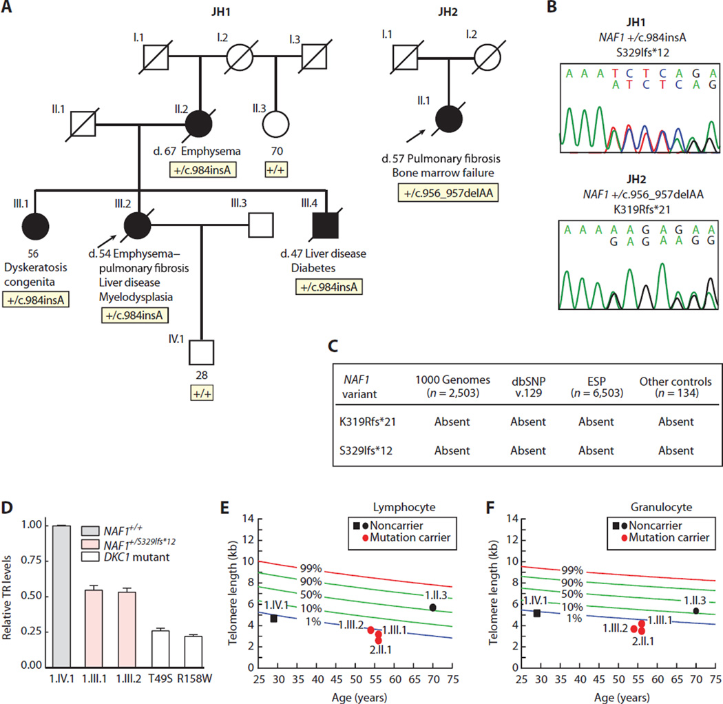
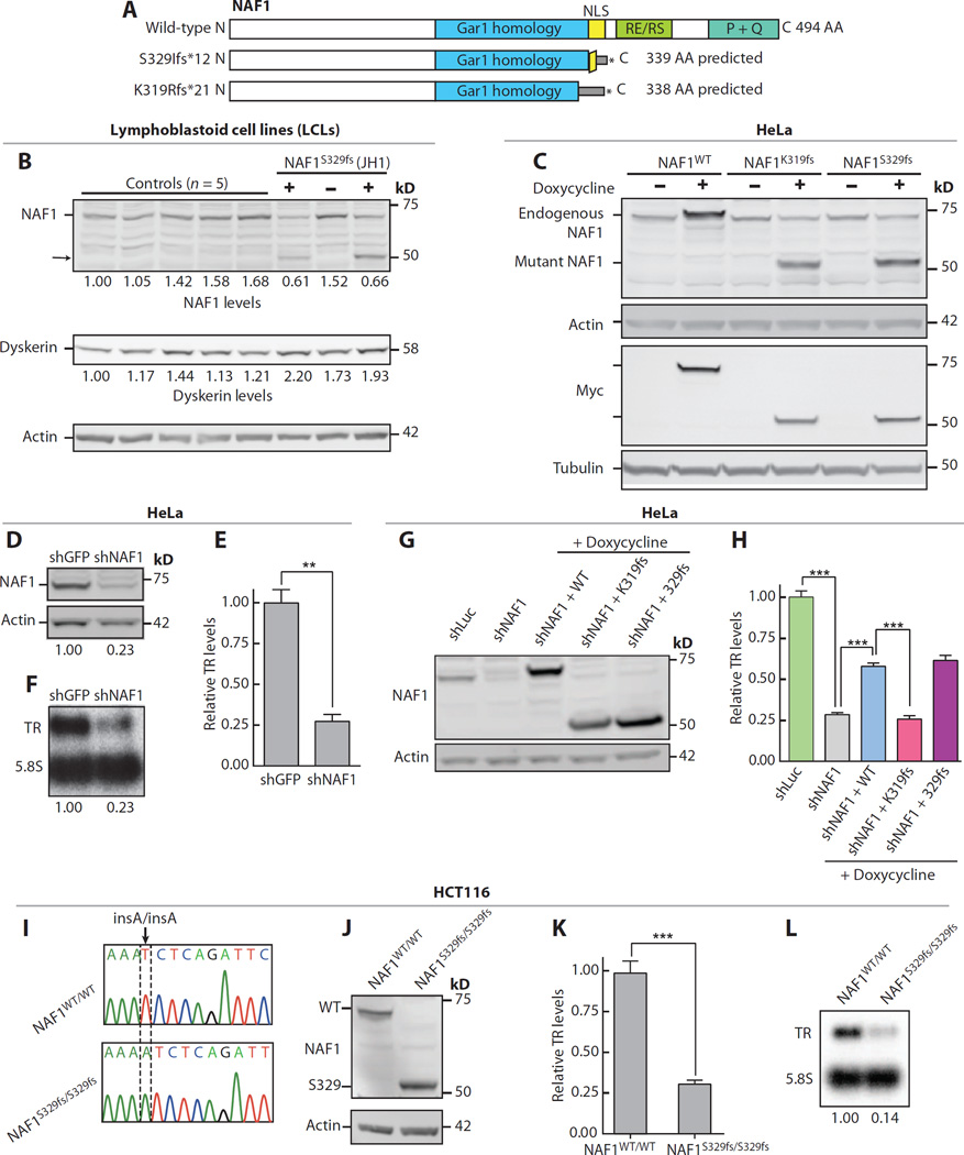
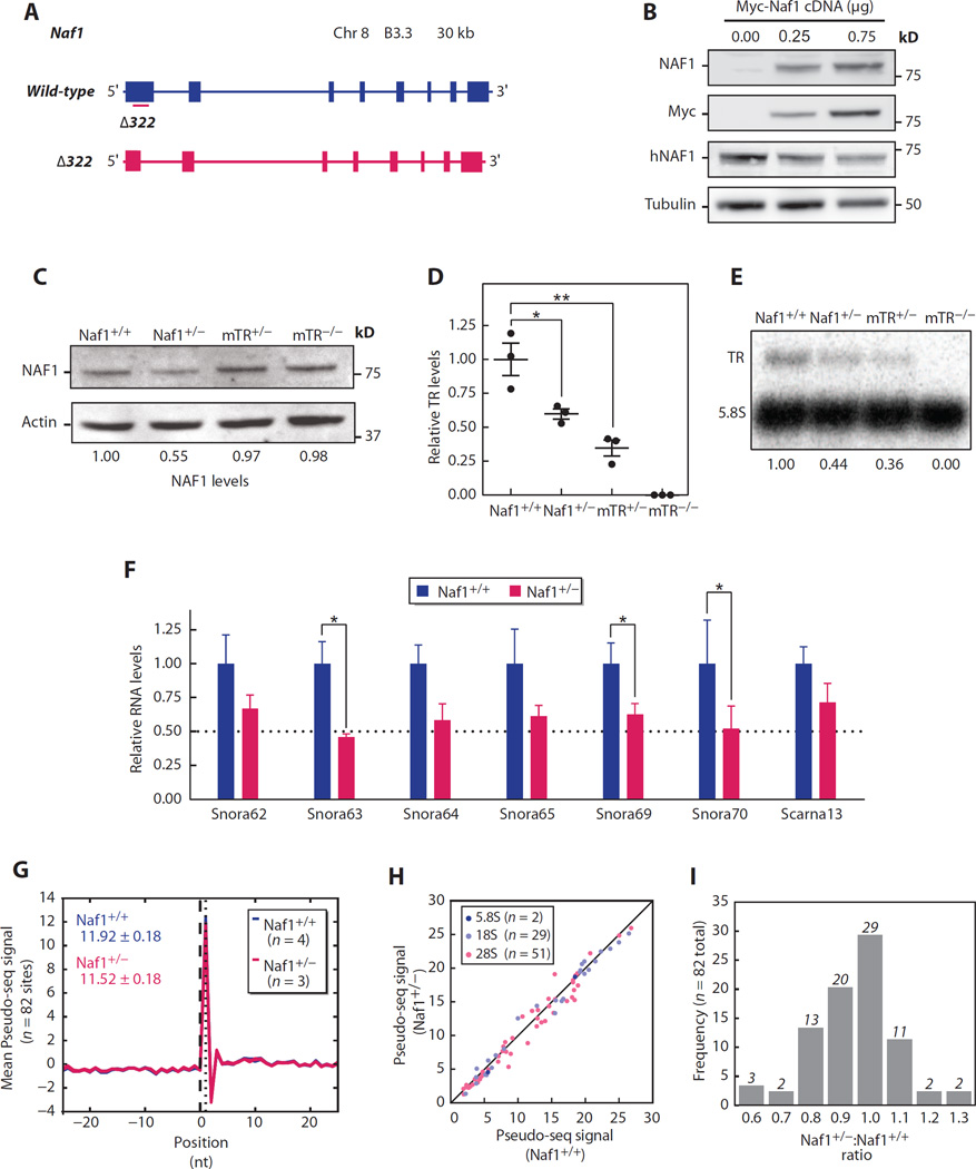
Chronic obstructive pulmonary disease and pulmonary fibrosis have been hypothesized to represent premature aging phenotypes. At times, they cluster in families, but the genetic basis is not understood. We identified rare, frameshift mutations in the gene for nuclear assembly factor 1, NAF1, a box H/ACA RNA biogenesis factor, in pulmonary fibrosis-emphysema patients. The mutations segregated with short telomere length, low telomerase RNA levels, and extrapulmonary manifestations including myelodysplastic syndrome and liver disease. A truncated NAF1 was detected in cells derived from patients, and, in cells in which the frameshift mutation was introduced by genome editing, telomerase RNA levels were reduced. The mutant NAF1 lacked a conserved carboxyl-terminal motif, which we show is required for nuclear localization. To understand the disease mechanism, we used CRISPR (clustered regularly interspaced short palindromic repeats)/Cas9 (CRISPR-associated protein-9 nuclease) to generate Naf1(+/-) mice and found that they had half the levels of telomerase RNA. Other box H/ACA RNA levels were also decreased, but rRNA pseudouridylation, which is guided by snoRNAs, was intact. Moreover, first-generation Naf1(+/-) mice showed no evidence of ribosomal pathology. Our data indicate that disease in NAF1 mutation carriers is telomere-mediated; they show that NAF1 haploinsufficiency selectively disturbs telomere length homeostasis by decreasing the levels of telomerase RNA while sparing rRNA pseudouridylation.


Figures

Fig. 1
Fig. 1. Rare NAF1 variants segregate with short telomere disease phenotypes and low TR levels
(A) Pedigrees of cases identified in a Johns Hopkins–based Registry (JH1 and JH2) with NAF1 genotypes shown below the individuals sequenced. NLS, nuclear localization signal. (B) Chromatograms of rare NAF1 variants by Sanger sequencing. (C) NAF1 variants are absent in public databases, as well as additional controls that we sequenced. dbSNP, Single Nucleotide Polymorphism Database; ESP, Exome Sequencing Project. (D) TR levels by qRT-PCR in LCLs with pedigree identifiers referring to (A). TR levels from DKC1 mutation carriers serve as a positive control, and the DKC1 missense mutations are annotated below. Means are from three separate RNA isolations and experiments, and the error bars represent SEM. (E and F) Age-adjusted telomere length in lymphocytes and granulocytes, respectively, measured by flow cytometry and fluorescence in situ hybridization (flow-FISH), with pedigree identifiers corresponding to individuals in (A). The nomogram was constructed from 192 healthy controls, and the calculated percentiles are labeled.
Fig. 2
Fig. 2. Disease-associated NAF1 mutations disturb TR stability
(A) Schema of NAF1 protein showing its conserved domains and predicted size of the truncated proteins (NP_612395.2). AA, amino acid. (B) Western blot of NAF1 and dyskerin in LCLs derived from unrelated controls and individuals from JH1 family. The “+” refers to NAF1 S329fs heterozygous mutation carriers (1.III.1 and I.III.2), and “−” refers to a noncarrier family member (I.IV.1). Pedigree identifiers refer to Fig. 1A. (C)Western blot for Myc-tagged NAF1 at an isogenic doxycycline-inducible locus in HeLa cells. (D) Immunoblot of NAF1 in HeLa cells after shRNA knockdown (day 8 time point). (E and F) TR levels measured by qRT-PCR and Northern blot, respectively. Mean for qRT-PCR result represents three independent experiments (P = 0.001). (G) Immunoblot for NAF1 after stable shRNA knockdown in HeLa cells with doxycycline induction of Myc-tagged NAF1. (H) TR levels measured by qRT-PCR after stable shRNA knockdown (48 hours after induction). Mean shown is from three technical replicates. P value for shLuc versus shNAF is <0.0001, for shNAF1 versus shNAF1 + WT (wild type) is 0.0002, and for shNAF1 + WT versus shNAF1 + K319fs is 0.0003. (I) Chromatogram of homozygous c.984insA S329fs disease-associated mutation knocked into the endogenous NAF1 locus of HCT116 cells using CRISPR/Cas9. (J) NAF1 immunoblot on lysates from CRISPR/Cas9-edited HCT116 cells. (K and L) TR levels quantified by qRT-PCR and Northern blot, respectively. For (K), mean reflects four independent RNA isolations and P = 0.0001. Error bars represent SEM. **P < 0.01 and ***P < 0.001 (Student’s t test).
Fig. 3
Fig. 3. Mutant NAF1 lacks a C-terminal NLS
(A) NAF1 amino acid alignment within and flanking the NLS for disease-associated mutations and those studied for nuclear localization by immunofluorescence are additionally shown. The four conserved lysine residues are indicated by dots above, and the bipartite NLS sequences are underlined. (B) Western blot of Myc-tagged NAF1 in the HeLa doxycycline-inducible system after 3-hour exposure to leptomycin B. (C) Immunofluorescence images showing subcellular localization of Myc-tagged NAF1 (green). Fluorophores in the three-color overlay are labeled to the left. Images were taken at ×63 magnification (scale bar, 20 µm). DAPI, 4′,6-diamidino-2-phenylindole. (D) Quantification of the NAF1 cytoplasmic fraction represents the intensity of cytoplasmic anti-Myc staining (defined by tubulin-positive area) relative to the nuclear staining (DAPI area). About 100 cells were quantified for each cell line, and mean values are shown with error bars representing SEM. Analysis was performed blinded to genotype. (E)Western blot of total (T), cytoplasmic (C), and nuclear (N) protein lysates of parental HCT116 and CRISPR/Cas9-edited HCT116 cells. For the quantification shown below, the total and cytoplasmic fractions were normalized to glyceraldehyde phosphate dehydrogenase (GAPDH), whereas the nuclear fraction was normalized to poly(ADP-ribose) polymerase (PARP). ***P < 0.001 (Student’s t test).
Fig. 4
Fig. 4. Naf1+/− mice have low TR and snoRNA levels, but rRNA pseudouridylation is intact
(A) Schema of wild-type Naf1 locus and CRISPR/Cas9-induced 322-bp deletion (Δ322) in exon 1. (B) Immunoblot of lysate from human embryonic kidney (HEK) 293FT cells transfected with Myc-tagged Naf1 cDNA-containing plasmid at shown concentrations. Immunoblot for human NAF1 (hNAF1) and mouse NAF1 and Myc shows specificity of the murine NAF1 antibody. (C) Immunoblot for NAF1 in adult skin fibroblasts. (D) TR measured by qRT-PCR (total spleen RNA, n = 3 mice per group) and normalized to hypoxanthine-guanine phosphoribosyltransferase (Hprt). For Naf1+/+ versus Naf1+/− comparison, P = 0.03; for Naf1+/+ versus mTR+/− comparison, P = 0.008. (E) Northern blot for mTR (mouse TR) normalized to 5.8S. (F) H/ACA RNA levels measured by qRT-PCR (n = 3 to 8 mice per group, total spleen RNA). P values for the comparisons for Snora63, Snora69, and Snora70 are 0.030, 0.048, and 0.036, respectively. (G) Average Pseudo-seq signal for 82 detected rRNA Ψ positions and surrounding 51-nucleotide window are shown in this metaPsi plot derived for Naf1+/+ (n = 4, blue) and Naf1+/− (n = 3, red) mice. The plot includes seven lines in total. Mean values are shown in the left upper corner. (H) Scatter plot of average Pseudo-seq signal ratio in Naf1+/+ versus Naf1+/− mice graphed for 82 sites in three rRNA subunits. (I) Frequency distribution of Pseudo-seq signal ratio in Naf1+/− compared with Naf1+/+ mice. Data are expressed as means ± SEM. *P < 0.05 and **P < 0.01 (Student’s t test).

Similar articles

Cited by

References

    1. Xu J, Murphy SL, Kochanek KD, Bastian BA. Deaths: Final data for 2013. Natl. Vital Stat. Rep. 2016;64:1–119. - PubMed
    1. Stanley SE, Armanios M. The short and long telomere syndromes: Paired paradigms for molecular medicine. Curr. Opin. Genet. Dev. 2015;33:1–9. - PMC - PubMed
    1. Stanley SE, Chen JJL, Podlevsky JD, Alder JK, Hansel NN, Mathias RA, Qi X, Rafaels NM, Wise RA, Silverman EK, Barnes KC, Armanios M. Telomerase mutations in smokers with severe emphysema. J. Clin. Invest. 2015;125:563–570. - PMC - PubMed
    1. Steele MP, Speer MC, Loyd JE, Brown KK, Herron A, Slifer SH, Burch LH, Wahidi MM, Phillips JA, III, Sporn TA, McAdams HP, Schwarz MI, Schwartz DA. Clinical and pathologic features of familial interstitial pneumonia. Am. J. Respir. Crit. Care Med. 2005;172:1146–1152. - PMC - PubMed
    1. Silverman EK, Weiss ST, Drazen JM, Chapman HA, Carey V, Campbell EJ, Denish P, Silverman RA, Celedon JC, Reilly JJ, Ginns LC, Speizer FE. Gender-related differences in severe, early-onset chronic obstructive pulmonary disease. Am. J. Respir. Crit. Care Med. 2000;162:2152–2158. - PubMed