. 2016 Dec;37(12):4472-4486.
doi: 10.1002/hbm.23322. Epub 2016 Aug 11.

Functional dissociation between anterior temporal lobe and inferior frontal gyrus in the processing of dynamic body expressions: Insights from behavioral variant frontotemporal dementia

Affiliations

Functional dissociation between anterior temporal lobe and inferior frontal gyrus in the processing of dynamic body expressions: Insights from behavioral variant frontotemporal dementia

Jan Jastorff et al. Hum Brain Mapp. 2016 Dec.

Abstract

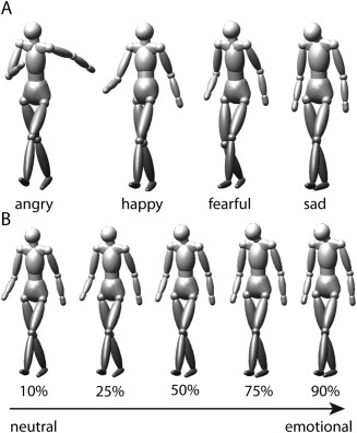
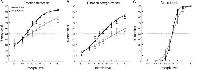
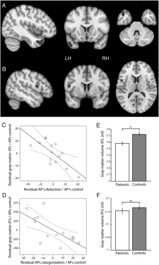
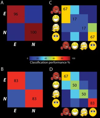
Several brain regions are involved in the processing of emotional stimuli, however, the contribution of specific regions to emotion perception is still under debate. To investigate this issue, we combined behavioral testing, structural and resting state imaging in patients diagnosed with behavioral variant frontotemporal dementia (bvFTD) and age matched controls, with task-based functional imaging in young, healthy volunteers. As expected, bvFTD patients were impaired in emotion detection as well as emotion categorization tasks, testing dynamic emotional body expressions as stimuli. Interestingly, their performance in the two tasks correlated with gray matter volume in two distinct brain regions, the left anterior temporal lobe for emotion detection and the left inferior frontal gyrus (IFG) for emotion categorization. Confirming this observation, multivoxel pattern analysis in healthy volunteers demonstrated that both ROIs contained information for emotion detection, but that emotion categorization was only possible from the pattern in the IFG. Furthermore, functional connectivity analysis showed reduced connectivity between the two regions in bvFTD patients. Our results illustrate that the mentalizing network and the action observation network perform distinct tasks during emotion processing. In bvFTD, communication between the networks is reduced, indicating one possible cause underlying the behavioral symptoms. Hum Brain Mapp 37:4472-4486, 2016. © 2016 Wiley Periodicals, Inc.

Keywords: connectivity; emotion; frontotemporal dementia; functional imaging; mentalizing.


Figures

Figure 1
Figure 1
Stimuli. (A) Example frames taken from 4 prototypical stimuli displaying the emotions angry, happy, fearful, and sad used in the functional imaging experiment. (B) Illustration of the morphed stimuli indicating different morph levels between neutral and emotional (sad) gaits tested during the behavioral experiment.
Figure 2
Figure 2
Comparison of gray matter volumes in bvFTD patients and controls. Shown in yellow are significantly atrophic voxels in the bvFTD group (P < 0.001 uncorrected at P < 0.05 FWE cluster size correction). Red voxels indicate the regions involved in emotion processing as defined in Jastorff et al. [2015], containing significantly less gray matter in patients compared to controls. [Color figure can be viewed at http://wileyonlinelibrary.com]
Figure 3
Figure 3
Behavioral performance. Percent emotional responses (± SEM) as a function of morph level for the emotion detection task (A), the emotion categorization task (B), and the control task (C). Solid lines indicate the fit of the psychometric curve.
Figure 4
Figure 4
Brain regions showing a significant correlation between (A) patients' APs in the emotion detection task and gray matter volume and (B) patients' APs in the emotion categorization task and gray matter volume. Volumes are shown at P < 0.01 uncorrected for illustration purposes. (C) Partial correlation between gray matter volume of the ATL ROI and the detection APs, when correcting for the influence of the APs obtained in the control task. (D) Partial correlation between gray matter volume of the IFG ROI and the categorization APs, when correcting for the influence of the APs obtained in the control task. (E) Comparison of gray matter volume in the ATL ROI (P < 0.01) between patients and controls. (F) Comparison of gray matter volume in the IFG ROI (P = 0.33) between patients and controls.
Figure 5
Figure 5
Resting state analysis. Red and blue spheres illustrate the ATL and IFG ROIs shown in Figure 4. Red and blue voxels exhibit significantly (P < 0.001 uncorrected at P < 0.05 FWE cluster size correction) reduced functional connectivity between the ATL ROI and the rest of the brain, and the IFG ROI and the rest of the brain, respectively. [Color figure can be viewed at http://wileyonlinelibrary.com]
Figure 6
Figure 6
Classification performance levels of the ATL ROI (A, C) and the IFG ROI (B, D) discriminating emotional and neutral stimuli and identifying the four emotions in young healthy volunteers. [Color figure can be viewed at http://wileyonlinelibrary.com]

References

    1. Abdollahi RO, Jastorff J, Orban GA (2013): Common and segregated processing of observed actions in human SPL. Cereb Cortex 23:2734–2753. - PubMed
    1. Amlerova J, Cavanna AE, Bradac O, Javurkova A, Raudenska J, Marusic P (2014): Emotion recognition and social cognition in temporal lobe epilepsy and the effect of epilepsy surgery. Epilepsy Behav 36:86–89. - PubMed
    1. Anwander A, Tittgemeyer M, von Cramon DY, Friederici AD, Knosche TR (2007): Connectivity‐Based Parcellation of Broca's Area. Cereb Cortex 17:816–825. - PubMed
    1. Baez S, Manes F, Huepe D, Torralva T, Fiorentino N, Richter F, Huepe‐Artigas D, Ferrari J, Montanes P, Reyes P, Matallana D, Vigliecca NS, Decety J, Ibanez A (2014): Primary empathy deficits in frontotemporal dementia. Front Aging Neurosci 6:262. - PMC - PubMed
    1. Barrett LF, Satpute AB (2013): Large‐scale brain networks in affective and social neuroscience: Towards an integrative functional architecture of the brain. Curr Opin Neurobiol 23:361–372. - PMC - PubMed

Publication types

MeSH terms