

Age-related differences in autism: The case of white matter microstructure
- PMID: 27511627
- PMCID: PMC6866893
- DOI: 10.1002/hbm.23345
Age-related differences in autism: The case of white matter microstructure
Abstract
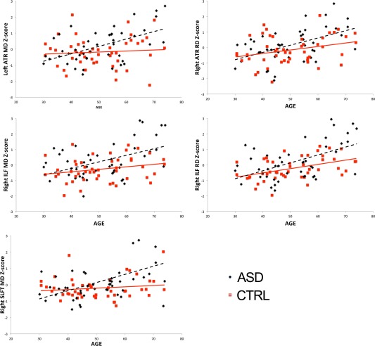
Autism spectrum disorder (ASD) is typified as a brain connectivity disorder in which white matter abnormalities are already present early on in life. However, it is unknown if and to which extent these abnormalities are hard-wired in (older) adults with ASD and how this interacts with age-related white matter changes as observed in typical aging. The aim of this first cross-sectional study in mid- and late-aged adults with ASD was to characterize white matter microstructure and its relationship with age. We utilized diffusion tensor imaging with head motion control in 48 adults with ASD and 48 age-matched controls (30-74 years), who also completed a Flanker task. Intra-individual variability of reaction times (IIVRT) measures based on performance on the Flanker interference task were used to assess IIVRT-white matter microstructure associations. We observed primarily higher mean and radial diffusivity in white matter microstructure in ASD, particularly in long-range fibers, which persisted after taking head motion into account. Importantly, group-by-age interactions revealed higher age-related mean and radial diffusivity in ASD, in projection and association fiber tracts. Subtle dissociations were observed in IIVRT-white matter microstructure relations between groups, with the IIVRT-white matter association pattern in ASD resembling observations in cognitive aging. The observed white matter microstructure differences are lending support to the structural underconnectivity hypothesis in ASD. These reductions seem to have behavioral percussions given the atypical relationship with IIVRT. Taken together, the current results may indicate different age-related patterns of white matter microstructure in adults with ASD. Hum Brain Mapp 38:82-96, 2017. © 2016 Wiley Periodicals, Inc.
Keywords: DTI; adults; autism; interference control; intra-individual variability; underconnectivity; white matter.
© 2016 Wiley Periodicals, Inc.
Figures
References
-
- Ameis SH, Catani M (2015): Altered white matter connectivity as a neural substrate for social impairment in Autism Spectrum Disorder. Cortex 62:158–181. - PubMed
-
- American Psychiatric Association. (2000) Diagnostic and Statistical Manual of Mental Disorders, 4th ed., text rev. Washington DC: American Psychiatric Association. Arlington, VA, USA
-
- Andersson JL, Skare S, Ashburner J (2003): How to correct susceptibility distortions in spin‐echo echo‐planar images: Application to diffusion tensor imaging. Neuroimage 20:870–888. - PubMed
Publication types
MeSH terms
LinkOut - more resources
Full Text Sources
Other Literature Sources
Medical
