. 2016 Oct 15;214(suppl 3):S319-S325.
doi: 10.1093/infdis/jiw270. Epub 2016 Aug 10.

FAM134B, the Selective Autophagy Receptor for Endoplasmic Reticulum Turnover, Inhibits Replication of Ebola Virus Strains Makona and Mayinga

Affiliations

FAM134B, the Selective Autophagy Receptor for Endoplasmic Reticulum Turnover, Inhibits Replication of Ebola Virus Strains Makona and Mayinga

Abhilash I Chiramel et al. J Infect Dis. .

Abstract

Selective autophagy of the endoplasmic reticulum (termed ER-phagy) is controlled by members of the FAM134 reticulon protein family. Here we used mouse embryonic fibroblasts from mice deficient in FAM134B to examine the role of the ER in replication of historic (Mayinga) or contemporary (Makona GCO7) strains of Ebola virus (EBOV). Loss of FAM134B resulted in 1-2 log10 higher production of infectious EBOV, which was associated with increased production of viral proteins GP and VP40 and greater accumulation of nucleocaspid lattices. In addition, only 10% of wild-type cells contained detectable nucleoprotein, whereas knockout of FAM134B resulted in 80% of cells positive for nucleoprotein. Together, these data suggest that FAM134B-dependent ER-phagy is an important limiting event in EBOV replication in mouse cells and may have implications for further development of antiviral therapeutics and murine models of infection.

Keywords: ER-phagy; Ebolavirus; FAM134B; mouse models; reticulon; selective autophagy; virus replication.


Figures

Figure 1.
Figure 1.
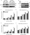
FAM134B restricts replication of Ebola virus (EBOV) in mouse embryonic fibroblasts (MEFs). A, Endogenous expression of FAM134B in MEFs. FAM134B+/+ (wild-type) or FAM134B−/− cells were cultured under complete medium (Dulbecco's modified Eagle's medium [DMEM]) or subjected to nutrient deprivation with Earl's balanced salt solution (EBSS) or Hank's buffered saline solution (HBSS) for 6 hours in the presence of dimethyl sulfoxide (DMSO) or bafilomycin A1 (BAFA1). FAM134B levels were analyzed by Western blots. B, FAM134B expression upon EBOV strain Makona GCO7 (EBOV–Makona GCO7) infection. FAM134B+/+ cells were infected with EBOV–Makona GCO7 at a multiplicity of infection (MOI) of 10 for 3 days. Cells were treated with vehicle or BAFA1 (100 nM) for 6 hours before samples were harvested. FAM134B expression was analyzed by Western blots. CF, FAM134B+/+ or FAM134B−/− MEFs were infected with EBOV-Mayinga or EBOV–Makona GCO7 at MOIs of 0.01 or 1, and supernatants were harvested 1, 3, 5, and 7 days after infection for titration. Error bars represent mean ± SD of experimental triplicates. Data are representative of 1 of 2 experiments. *P < .05 and ****P < .0001, by 2-way analysis of variance with the Sidak multiple comparison test. Abbreviation: FFU, focus-forming units.
Figure 2.
Figure 2.
Expression of VP40 (A) and glycoprotein (GP; B) in FAM134B+/+ or FAM134B−/− cells. MEFs were infected with Ebola virus strain Mayinga (EBOV-Mayinga) or EBOV–Makona GCO7 at a multiplicity of infection (MOI) of 1. Visualization of actin or vinculin (for high-molecular-weight proteins) were used as loading controls. C, Nucleoprotein (NP) staining in EBOV-Makona–infected FAM134B+/+ or FAM134B−/− MEFs. Cells were infected with EBOV-Makona at a MOI of 1 for 6 days. Samples were stained for NP (green) and nuclei (DAPI; blue), and images were obtained by confocal microscopy. D, Quantification of infected NP-positive cells.
Figure 3.
Figure 3.
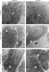
Ultrastructural analysis of FAM134B+/+ or FAM134B−/− cells uninfected or infected with Ebola virus strain Mayinga (EBOV-Mayinga) or EBOV–Makona GCO7 at a multiplicity of infection of 1. Cells were fixed with 2.5% glutaraldehyde 3 days after infection and processed for transmission electron microscopy. Subcellular localization of endoplasmic reticulum (ER) (white arrows) and cytoplasmic nucleocapsid accumulation (white arrowheads) is indicated. Scale bar, 2 µm.

References

    1. Bah EI, Lamah MC, Fletcher T et al. . Clinical presentation of patients with Ebola virus disease in Conakry, Guinea. N Engl J Med 2015; 372:40–7. - PubMed
    1. de La Vega MA, Stein D, Kobinger GP. Ebolavirus evolution: past and present. PLoS Pathog 2015; 11:e1005221. - PMC - PubMed
    1. Schieffelin JS, Shaffer JG, Goba A et al. . Clinical illness and outcomes in patients with Ebola in Sierra Leone. N Engl J Med 2014; 371:2092–100. - PMC - PubMed
    1. Marzi A, Feldmann F, Hanley PW, Scott DP, Gunther S, Feldmann H. Delayed disease progression in cynomolgus macaques infected with Ebola virus makona strain. Emerg Infect Dis 2015; 21:1777–83. - PMC - PubMed
    1. Chan SY, Ma MC, Goldsmith MA. Differential induction of cellular detachment by envelope glycoproteins of Marburg and Ebola (Zaire) viruses. J Gen Virol 2000; 81:2155–9. - PubMed

Publication types