. 2016 Aug 9;8(4):1759091416662808.
doi: 10.1177/1759091416662808. Print 2016 Aug.

Diffusible Factors Secreted by Glioblastoma and Medulloblastoma Cells Induce Oxidative Stress in Bystander Neural Stem Progenitors

Affiliations

Diffusible Factors Secreted by Glioblastoma and Medulloblastoma Cells Induce Oxidative Stress in Bystander Neural Stem Progenitors

Neha Sharma et al. ASN Neuro. .

Abstract

Harmful effects that alter the homeostasis of neural stem or progenitor cells (NSPs) can affect regenerative processes in the central nervous system. We investigated the effect of soluble factors secreted by control or (137)Cs-γ-irradiated glioblastoma or medulloblastoma cells on redox-modulated endpoints in recipient human NSPs. Growth medium harvested from the nonirradiated brain tumor cells, following 24 h of growth, induced prominent oxidative stress in recipient NSPs as judged by overall increases in mitochondrial superoxide radical levels (p < .001), activation of c-jun N-terminal kinase, and decrease in the active form of FoxO3a. The induced oxidative stress was associated with phosphorylation of p53 on serine 15, a marker of DNA damage, induction of the cyclin-cyclin dependent kinase inhibitors p21(Waf1) and p27(Kip1), and perturbations in cell cycle progression (p < .001). These changes were also associated with increased apoptosis as determined by enhanced annexin V staining (p < .001) and caspase 8 activation (p < .05) and altered expression of critical regulators of self-renewal, proliferation, and differentiation. Exposure of the tumor cells to radiation only slightly altered the induced oxidative changes in the bystander NSPs, except for medium from irradiated medulloblastoma cells that was more potent at inducing apoptosis in the NSPs than medium from nonirradiated cells (p < .001). The elucidation of such stressful bystander effects provides avenues to understand the biochemical events underlying the development or exacerbation of degenerative outcomes associated with brain cancers. It is also relevant to tissue culture protocols whereby growth medium conditioned by tumor cells is often used to support the growth of stem cells.

Keywords: brain tumors; intercellular communication; ionizing radiation or bystander effect; neural stem progenitors; reactive oxygen species.


Figures

Figure 1.
Figure 1.
Factors secreted by brain tumor cells modulate superoxide levels in mitochondria of bystander NSPs. H9 NSPs cultured for 24 h in medium harvested from control (CCM) or irradiated (ICM) T98G (glioblastoma) or Daoy (medulloblastoma) cell cultures were subjected to MitoSox analysis using flow cytometry. Bar graph represents average median fluorescence intensity in arbitrary units (au) from three independent experiments ± SEM. Values in the different experimental conditions were compared using a one-way ANOVA, with the Tukey test to control for multiple comparisons.
Figure 2.
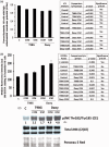
Figure 2.
Factors secreted by brain tumor cells alter mitochondrial membrane potential (ΔΨm), upregulate intracellular ROS levels, and activate redox-modulated kinase in bystander NSPs. (a) H9 NSPs cultured for 24 h with medium conditioned for 24 h by control (CCM) or irradiated (ICM) T98G or Daoy brain tumor cells were subjected to ΔΨm analysis using JC-1 probe by flow cytometry. Bar graph is average of three independent experiments ± SEM of NSPs with high ΔΨm. (b) H9 NSPs were treated as in (a) and subjected to analyses of intracellular ROS using CM-DCFDA assay by flow cytometry. Bar graph represents average of median fluorescence intensity in arbitrary units (au) ± SEM (n = 3). (c) H9 NSPs were treated as in (a) and subjected to immunoblot analysis (n = 3). One way ANOVA was used in analyses of the results in panel 2 A and χ2 test was used in analysis of the results in panel 2B as detailed in Materials and Methods section.
Figure 3.
Figure 3.
Factors secreted by brain tumor cells alter expression of regulatory genes involved in ROS detoxification in bystander NSPs. H9 NSPs cultured for 24 h with medium conditioned for 24 h by control (CCM) or irradiated (ICM) T98G or Daoy cell cultures were subjected to immunoblot analysis of lysates enriched in nuclear extract (NE) proteins (n = 3). The relative intensity (R.I.) is intensity (I) of a band (z) normalized against its control (c) and respective Ponceau S Red intensity (P). R.I. = [I(z)/P(z)]/[I(c)/P(c)].
Figure 4.
Figure 4.
Propagation of oxidative stress from brain tumor cells is associated with induction of DNA damage responsive genes in bystander NSPs. H9 NSPs cultured for 24 h in medium conditioned for 24 h by control (CCM) or irradiated (ICM) T98G or Daoy brain tumor cells were subjected to immunoblot analysis in lysates enriched in the nuclear fraction (n = 3). The relative intensity (R.I.) is intensity (I) of a band (z) normalized against its control (c) and respective Ponceau S Red intensity (P). R.I. = [I(z)/P(z)]/[I(c)/P(c)].
Figure 5.
Figure 5.
Propagation of oxidative stress from brain tumor cells to bystander NSPs is associated with perturbation in cell cycle progression. H9 NSPs cultured for 24 h in medium conditioned for 24 h by T98G or Daoy cells were subjected to cell cycle analysis by flow cytometry. Events (10,000) were collected for analysis and representative histograms are shown (ordinates are number of cells and abscissa is DNA content as assessed by propidium iodide staining; n = 3).
Figure 6.
Figure 6.
Factors secreted by brain tumor cells promote apoptotic death in bystander NSPs. (a) H9 NSPs cultured for 24 h in medium conditioned for 24 h by T98G or Daoy cells were subjected to Annexin V/PI assay and analyzed by flow cytometry. Events (10,000) were collected for the analysis and changes in cell proportions in response to the different experimental conditions were tested by the χ2 test, using the Holm-Sidak test to control for multiple comparisons. Significance was set at p < .05. Bar graph represents average of four independent experiments ± SEM. (b) H9 cells were treated as in (a) and subjected for caspase 8 assay by flow cytometry. Bar graph represents average median fluorescence intensity in arbitrary units (au) from five independent experiments ± SEM. The average values for each treatment were compared using a one-way ANOVA, with the Tukey test to control for multiple comparisons.
Figure 7.
Figure 7.
Factors secreted by brain tumor cells alter levels of survival regulators in bystander NSPs. (a) H9 NSPs cultured for 24 h in medium conditioned for 24 h by T98G or Daoy cells were subjected to immunoblot analysis in lysates enriched in either cytoplasmic (CE) or nuclear fractions (NE) (n = 3). (b) H9 NSPs were treated as in (a) and analysed by immunoblotting for LC3I lipidation to LC3II (n = 3). The relative intensity (R.I.) is intensity (I) of a band (z) normalized against its control (c) and respective Ponceau S Red intensity (P). R.I. = [I(z)/P(z)]/[I(c)/P(c)].

References

    1. Acharya M. M., Lan M. L., Kan V. H., Patel N. H., Giedzinski E., Tseng B. P., Limoli C. L. (2010) Consequences of ionizing radiation-induced damage in human neural stem cells. Free Radical Biology & Medicine 49: 1846–1855. - PubMed
    1. Aickin M., Gensler H. (1996) Adjusting for multiple testing when reporting research results: The Bonferroni vs Holm methods. American Journal of Public Health 86: 726–728. - PMC - PubMed
    1. Azzam E. I., De Toledo S. M., Harris A. L., Ivanov V., Zhou H., Amundson S. A., Hei T. K. (2013) The ionizing radiation-induced bystander effect: Evidence, mechanism and significance. In: Sonis S. T., Keefe D. M. (eds) Pathobiology of cancer regimen-related toxicities, New York, NY: Springer, pp. 42–68.
    1. Azzam E. I., de Toledo S. M., Little J. B. (2003) Oxidative metabolism, gap junctions and the ionizing radiation-induced bystander effect. Oncogene 22: 7050–7057. - PubMed
    1. Azzam E. I., Jay-Gerin J. P., Pain D. (2012) Ionizing radiation-induced metabolic oxidative stress and prolonged cell injury. Cancer Letters 327: 48–60. - PMC - PubMed

MeSH terms

LinkOut - more resources