Review
. 2016 Nov 1;116(5):2093-2104.
doi: 10.1152/jn.00221.2016. Epub 2016 Aug 10.

White matter and cognition: making the connection

Affiliations
Review

White matter and cognition: making the connection

Christopher M Filley et al. J Neurophysiol. .

Abstract

Whereas the cerebral cortex has long been regarded by neuroscientists as the major locus of cognitive function, the white matter of the brain is increasingly recognized as equally critical for cognition. White matter comprises half of the brain, has expanded more than gray matter in evolution, and forms an indispensable component of distributed neural networks that subserve neurobehavioral operations. White matter tracts mediate the essential connectivity by which human behavior is organized, working in concert with gray matter to enable the extraordinary repertoire of human cognitive capacities. In this review, we present evidence from behavioral neurology that white matter lesions regularly disturb cognition, consider the role of white matter in the physiology of distributed neural networks, develop the hypothesis that white matter dysfunction is relevant to neurodegenerative disorders, including Alzheimer's disease and the newly described entity chronic traumatic encephalopathy, and discuss emerging concepts regarding the prevention and treatment of cognitive dysfunction associated with white matter disorders. Investigation of the role of white matter in cognition has yielded many valuable insights and promises to expand understanding of normal brain structure and function, improve the treatment of many neurobehavioral disorders, and disclose new opportunities for research on many challenging problems facing medicine and society.

Keywords: cognition; dementia; glial cells; myelin; white matter.


Figures

Fig. 1.
Fig. 1.
Axial fluid-attenuated inversion recovery (FLAIR) MRI scans showing the scattered white matter lesions of leukoaraiosis (A) and confluent white matter hyperintensity (B) consistent with Binswanger's disease.
Fig. 2.
Fig. 2.
DTI scan of a normal adult brain showing 3 white matter tracts. Color coding permits the demonstration of tracts oriented within the right-left, anterior-posterior, and superior-inferior planes: red indicates the corpus callosum, green represents the arcuate fasciculus, and blue depicts the corticospinal tract. DTI image provided by M. Brown, Univ. of Colorado.
Fig. 3.
Fig. 3.
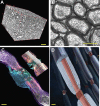
White matter structure. A: ∼40 μm × 60 μm × 60 μm volume of white matter (mouse optic nerve) reconstructed from serial block face electron microscopy, showing the composition of axons, myelin, astrocytes, oligodendrocytes, and vasculature. Changes in any of these components or in the tortuosity of the fibers will influence diffusional MRI, where the voxel volume is 100 μm × 100 μm × 100 μm in high-resolution MRI of rodents and typically 2 mm × 2 mm × 2 mm in human brain imaging. B: optic nerve axons in cross section showing multilaminar wrapping of compact myelin. C: 3-dimensional reconstruction of a node of Ranvier in mouse optic nerve from serial block face electron microscopy: myelin (purple), paranodal loops (salmon), perinodal astrocyte (green and blue). Inset: longitudinal slice through the node of Ranvier revealing the nodal gap (gray) between the paranodal loops of myelin containing high-density voltage-sensitive sodium channels. White, axon. D: illustration of myelinated axons, showing the multilaminar myelin sheath and electrogenic node of Ranvier. Scale bars: 10 μm in A, 1 μm in B–D.

References

    1. Abiraman K, Pol SU, O'Bara MA, Chen GD, Khaku ZM, Wang J, Thorn D, Vedia BH, Ekwegbalu EC, Li JX, Salvi RJ, Sim FJ. Anti-muscarinic adjunct therapy accelerates functional human oligodendrocyte repair. J Neurosci 35: 3676–3688, 2015. - PMC - PubMed
    1. Aguirre A, Dupree JL, Mangin JM, Gallo V. A functional role for EGFR signaling in myelination and remyelination. Nat Neurosci 10: 990–1002, 2007. - PubMed
    1. Albert ML, Feldman RG, Willis AL. The “subcortical dementia” of progressive supranuclear palsy. J Neurol Neurosurg Psychiatry 37: 121–130, 1974. - PMC - PubMed
    1. Alexander MP. Mild traumatic brain injury: pathophysiology, natural history, and clinical management. Neurology 45: 1253–1260, 1995. - PubMed
    1. Anderson VC, Obayashi JT, Kaye JA, Quinn JF, Berryhill P, Riccelli LP, Peterson D, Rooney WD. Longitudinal relaxographic imaging of white matter hyperintensities in the elderly. Fluids Barriers CNS 11: 24, 2014. - PMC - PubMed

Publication types