. 2016 Oct 14;90(21):9582-9597.
doi: 10.1128/JVI.01241-16. Print 2016 Nov 1.

Baculovirus FP25K Localization: Role of the Coiled-Coil Domain

Affiliations

Baculovirus FP25K Localization: Role of the Coiled-Coil Domain

Tyler A Garretson et al. J Virol. .

Abstract

Two types of viruses are produced during the baculovirus life cycle: budded virus (BV) and occlusion-derived virus (ODV). A particular baculovirus protein, FP25K, is involved in the switch from BV to ODV production. Previously, FP25K from the model alphabaculovirus Autographa californica multiple nucleopolyhedrovirus (AcMNPV) was shown to traffic ODV envelope proteins. However, FP25K localization and the domains involved are inconclusive. Here we used a quantitative approach to study FP25K subcellular localization during infection using an AcMNPV bacmid virus that produces a functional AcMNPV FP25K-green fluorescent protein (GFP) fusion protein. During cell infection, FP25K-GFP localized primarily to the cytoplasm, particularly amorphous structures, with a small fraction being localized in the nucleus. To investigate the sequences involved in FP25K localization, an alignment of baculovirus FP25K sequences revealed that the N-terminal putative coiled-coil domain is present in all alphabaculoviruses but absent in betabaculoviruses. Structural prediction indicated a strong relatedness of AcMNPV FP25K to long interspersed element 1 (LINE-1) open reading frame 1 protein (ORF1p), which contains an N-terminal coiled-coil domain responsible for cytoplasmic retention. Point mutations and deletions of this domain lead to a change in AcMNPV FP25K localization from cytoplasmic to nuclear. The coiled-coil and C-terminal deletion viruses increased BV production. Furthermore, a betabaculovirus FP25K protein lacking this N-terminal coiled-coil domain localized predominantly to the nucleus and exhibited increased BV production. These data suggest that the acquisition of this N-terminal coiled-coil domain in FP25K is important for the evolution of alphabaculoviruses. Moreover, with the divergence of preocclusion nuclear membrane breakdown in betabaculoviruses and membrane integrity in alphabaculoviruses, this domain represents an alphabaculovirus adaptation for nuclear trafficking of occlusion-associated proteins.

Importance: Baculovirus infection produces two forms of viruses: BV and ODV. Manufacturing of ODV involves trafficking of envelope proteins to the inner nuclear membrane, mediated partly through the FP25K protein. Since FP25K is present in alpha-, beta-, and gammabaculoviruses, it is uncertain if this trafficking function is conserved. In this study, we looked at alpha- and betabaculovirus FP25K trafficking by its localization. Alphabaculovirus FP25K localized primarily to the cytoplasm, whereas betabaculovirus FP25K localized to the nucleus. We found that an N-terminal coiled-coil domain present in all alphabaculovirus FP25K proteins, but absent in betabaculovirus FP25K, was critical for alphabaculovirus FP25K cytoplasmic localization. We believe that this represents an evolutionary process that partly led to the gain of function of this N-terminal coiled-coil domain in alphabaculovirus FP25K to aid in nuclear trafficking of occlusion-associated proteins. Due to betabaculovirus breakdown of the nuclear membrane before occlusion, this function is not needed, and the domain was lost or never acquired.


Figures

FIG 1
FIG 1
Generation of an AcMNPV FP25K-GFP fusion protein for FP25K localization studies. (A) AcMNPV-based bacmid (AcBacmid) transfection and subsequent continuous BV passaging in Sf21 cells lead to insertional mutagenesis of the fp25k locus. (Left) PCR of the fp25k locus from AcBacmid passages. Purified AcBacmid DNA from bacteria was transfected, and BVs from prior infections were used as the inoculum for sequential passaging in Sf21 cells 10 times. BV DNA was extracted, and the fp25k locus was amplified by PCR using fp25k-specific primers (Table 1). (Right) PCR confirmation of an fp25k mutant AcBacmid (AcBac-fp25k::287) clone. BV DNA from AcBacmid passage 10 was transformed into E. coli cells. A particular fp25k mutant clone (C9) was isolated, and the fp25k locus was amplified by PCR using the same fp25k-specific primers. NTC, nontemplate control. (B) Diagram of AcFP-GFP(ProPH) virus construction. All subsequent figures depict a similar construction utilizing E. coli DH10Bac-fp25k::287 cells. AcMNPV fp25k and gfp coding sequences were fused and introduced into the donor vector pFastBac1. The resulting pFB1-AcFP-GFP(ProPH) plasmid was transformed into E. coli DH10Bac-fp25k::287 cells. This cell line also contains the pMON7124 helper plasmid that encodes the Tn7 transposase allowing recombination between the mini-Tn7 attachment sites on bacmid bMON14272 at the polyhedrin (polh) locus and pFastBac1 (boxed). Bacmid bMON14272 in this cell-specific line contains the fp25k insertional mutant generated as described above for panel A. ProPH, polh promoter; L, linker sequence between fp25k and gfp CDSs. (C) Detection of the full-length AcMNPV FP25K-GFP fusion protein from AcFP-GFP(ProPH)-infected Tn5 and Sf9 cells by Western blotting. Tn5 and Sf9 cells were infected with the unfused AcBacGFP control virus and AcFP-GFP(ProPH). At 48 hpi, cell lysates were subjected to Western blot analysis using anti-GFP polyclonal antibody. (D) AcMNPV FP25K-GFP shows functionality in terms of reduced BV production during AcFP-GFP(ProPH) infection in the budding assay. Tn5 cells were infected with AcBacGFP (wild-type control), AcBac-fp25k::287-GFP (mutant), and AcFP-GFP(ProFP) at an MOI of 0.1. At different time points postinfection, BV was harvested, and titers were determined by qPCR. Statistical significances were determined by using the Student t test and are indicated by asterisks (*, P < 0.05; **, P < 0.005).
FIG 2
FIG 2
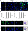
AcMNPV FP25K localizes primarily to the cytoplasm, with a small percentage being present in the nucleus. (A) Confocal microscopy of Tn5 (a to e) and Sf9 (g to k) cells infected with AcFP-GFP(ProPH) and AcBacGFP viruses, as indicated, at an MOI of 1. At 12 hpi (a and g), 24 hpi (b and h), 36 hpi (c and i), and 48 hpi (d to f and j to l), AcFP-GFP(ProPH)- and AcBacGFP-infected Tn5 and Sf9 cells were treated with the permeable nuclear stain Hoechst 33342 (blue, nucleus), and confocal images were taken. The yellow insets from samples from AcFP-GFP(ProPH) infection at 48 hpi (d and j) are shown in panels e and k. White arrows indicate locations where GFP is present in the nucleus. White arrowheads designate the described amorphous structures. Bars, 25 μm (a to d, f, g to j, and l) and 10 μm (e and k). (B and C) Quantitative analysis of AcMNPV FP25K-GFP localization in the cytoplasm and nucleus by confocal microscopy of AcFP-GFP(ProPH)-infected Tn5 (B) and Sf9 (C) cells. The amounts of GFP are based on 30 GFP-positive cells from each infection. Values are reported as a percentage of the total cellular AcMNPV FP25K-GFP in either the cytoplasm or the nucleus. N/A signifies that no AcMNPV FP25K-GFP was found at 12 hpi. Error bars represent the standard deviations of data from the 30 cells analyzed. Statistical significance is indicated by asterisks (**, P < 0.005). (D) z-stack analysis of Tn5 (a to e) and Sf9 (g to k) cells infected with AcFP-GFP(ProPH) virus at an MOI of 1. In the z axis, images were taken every 3 μm for Tn5-infected cells and every 2 μm for Sf9-infected cells. Cells were treated with the nuclear stain Hoechst 33342 (blue, nucleus). White arrows indicate GFP in the nucleus. The relative depth of the image in micrometers is indicated on top of each image. Bars, 10 μm. Panels f and l show schematics summarizing data from all the images taken in the z plane. The red lines indicate where the images were taken on the z axis.
FIG 3
FIG 3
polh and fp25k promoter-driven expression does not alter AcMNPV FP25K-GFP localization. (A) Diagrams indicating the differences between the AcFP-GFP(ProPH) and AcFP-GFP(ProFP) viruses. Both viruses were constructed as described in the legend of Fig. 1. However, for AcFP-GFP(ProFP), the AcMNPV fp25k coding sequence (in gray) containing its respective promoter sequence upstream (in light gray) was fused to gfp. ProPH, polh promoter; ProFP, fp25k promoter; L, linker sequence between fp25k and gfp CDSs. (B) Confirmation of lower AcMNPV FP25K-GFP expression levels in fp25k than in polh promoter-driven virus. Tn5 and Sf9 cells were infected with AcFP-GFP(ProFP) (gray bars) and AcFP-GFP(ProPH) (black bars) viruses at an MOI of 1. The level of GFP expression was determined at 48 hpi and is represented by the change in fluorescence units (ΔFU) in the infected sample relative to the uninfected sample. (C) Confocal microscopy at 48 hpi of Tn5 cells infected with AcFP-GFP(ProPH) and AcFP-GFP(ProFP) viruses at an MOI of 1. Cells were treated with the nuclear stain Hoechst 33342 (blue, nucleus). The yellow insets from the 48-hpi samples (a and c) (bar = 25 μm) are shown in panels b and d (bar = 10 μm). White arrows indicate locations where GFP is present in the nucleus. White arrowheads designate the described amorphous structures. (D) Quantitative analysis of AcMNPV FP25K-GFP localization in the cytoplasm and nucleus by confocal microscopy of AcFP-GFP(ProPH)- and AcFP-GFP(ProFP)-infected cells. The amounts of GFP were based on 30 GFP-positive cells from each infection. Values are reported as a percentage of the total cellular AcMNPV FP25K-GFP level in either the cytoplasm or nucleus. Error bars represent the standard deviations of data from the 30 cells analyzed. Statistical significance is indicated by the asterisks (**, P < 0.005).
FIG 4
FIG 4
The N-terminal coiled-coil domain of FP25K is found in all alphabaculoviruses but is completely lacking in the betabaculoviruses. Alignment of the baculovirus FP25K amino acid sequences was performed by using CLC Main Workbench. The FP25K or ORF61 homologues were extracted from currently available Baculoviridae genomes. Shown is a representative wide view of the whole FP25K protein (A), and the coiled-coil domain is boxed (B). The alpha- and betabaculovirus genera are indicated, along with the predicted coiled-coil domain, nucleic acid binding domain, and actin-binding helix at the top. The amino acid positions according to the alignment are also designated.
FIG 5
FIG 5
Mutation and deletion of the N-terminal coiled-coil domain of AcMNPV FP25K alter its localization. (A) Diagrams and prediction of AcMNPV FP25K coiled-coil formation in each point mutation construct. (Top) Location of the point mutations within the N-terminal coiled-coil domain of the AcMNPV FP25K-GFP fusion protein. The red “X” indicates the position of the point mutation in each of the constructs. These constructs were introduced into the mutant bacmid, as described in the legend of Fig. 1. (Bottom) Graphical output for prediction of AcMNPV FP25K coiled-coil formation from the primary sequence of wild-type FP25K and the L36P and D22P mutants by using the COILS program (http://www.ch.embnet.org/software/COILS_form.html). The y axis represents the probability of coiled-coil formation obtained by the program, and the x axis shows the position of the residue. Strong, high peaks by window 14 (green), window 21 (blue), and window 28 (red) represent a high probability of coiled-coil formation. (B) Confocal microscopy at 48 hpi of Tn5 cells infected with AcFP-GFP(ProFP), L36P, or D22P virus at an MOI of 1. Cells were treated with the nuclear stain Hoechst 33342 (blue, nucleus). Bars, 25 μm. White arrows indicate locations where GFP is present in the nucleus. White arrowheads designate the described amorphous structures. Dotted white circles indicate the position of the nucleus in cells with a diffuse GFP localization. (C) Quantitative analysis of AcMNPV FP25K-GFP localization in the cytoplasm and nucleus by confocal microscopy of AcFP-GFP(ProFP), L36P, or D22P samples. Samples were based on 30 GFP-positive cells from each infection. Values are reported as a percentage of the total cellular AcMNPV FP25K-GFP level in either the cytoplasm or the nucleus. Error bars represent the standard deviations of data from the 30 cells analyzed. Different letters indicate statistical significance between the localizations of the constructs (P < 0.05). (D) Diagrams showing the location of the deletions within the AcMNPV FP25K-GFP fusion protein. The red squares indicate the position of the deletion in each of the constructs. (E) Confocal microscopy at 48 hpi of Tn5 cells infected with AcFP-GFP(ProFP), ΔL13:L44, ΔG52:L139, and ΔK142:S206 viruses at an MOI of 1, as described above for panel B. (F) Quantitative analysis of AcMNPV FP25K-GFP localization in the cytoplasm and nucleus by confocal microscopy of AcFP-GFP(ProFP), ΔL13:L44, ΔG52:L139, and ΔK142:S206 samples, as described above for panel C.
FIG 6
FIG 6
BV production is altered by deletion of the N-terminal coiled-coil domain and C-terminal region of AcMNPV FP25K. Tn5 cells were infected with AcBacGFP (wild-type control), AcBac-fp25k::287-GFP (mutant), AcFP-GFP(ProFP) (deletion background), ΔL13:L44, ΔG52:L139, and ΔK142:S206 viruses at an MOI of 0.1. At 72 hpi, BV was harvested, and titers were then determined by qPCR. Statistical significance is indicated by asterisks (*, P < 0.05).
FIG 7
FIG 7
PxGV FP25K localizes primarily to the nucleus. (A) Diagrams indicating the differences between AcFP-GFP(ProPH) and PxFP-GFP(ProPH) viruses. The fusion constructs were introduced into the mutant bacmid, as described in the legend of Fig. 1. In PxFP-GFP(ProPH), the PxGV fp25k coding sequence (which does not contain an N-terminal coiled-coil domain) was fused to gfp. L, linker sequence between fp25k and gfp CDSs. (B) Detection of the full-length PxGV FP25K-GFP fusion protein from PxFP-GFP(ProPH)-infected Tn5 cells by Western blotting. Tn5 cells were infected with the unfused AcBacGFP control virus and PxFP-GFP(ProPH). At 48 hpi, cell lysates were subjected to Western blot analysis using an anti-GFP polyclonal antibody. The GFP protein is 27 kDa. The predicted molecular mass of the PxGV FP25K-GFP fusion protein is 42 kDa. (C) Confocal microscopy at 48 hpi of Tn5 cells infected with AcFP-GFP(ProPH) and PxFP-GFP(ProPH) viruses at an MOI of 1. Cells were treated with the nuclear stain Hoechst 33342 (blue, nucleus). The yellow insets from the 48-hpi samples (a and c) (bar = 25 μm) are shown in panels b and d (bar = 10 μm). White arrows indicate locations where GFP is present in the nucleus. White arrowheads designate the described amorphous structures. Dotted white circles indicate the position of the nucleus in cells with a diffuse GFP localization. (D) Quantitative analysis of AcMNPV FP25K-GFP localization in the cytoplasm and nucleus by confocal microscopy of AcFP-GFP(ProPH)- and PxFP-GFP(ProPH)-infected samples. Samples were based on 30 GFP-positive cells from each infection. Values are reported as a percentage of the total cellular AcMNPV FP25K-GFP level in either the cytoplasm or the nucleus. Error bars represent the standard deviations of data from the 30 cells analyzed. Statistical significance is indicated by asterisks (**, P < 0.005).
FIG 8
FIG 8
PxGV FP25K does not restrict virus budding to wild-type levels. Tn5 cells were infected with AcBacGFP (wild-type control), AcBac-fp25k::287-GFP (mutant), AcFP-GFP(ProFP), and PxFP-GFP(ProFP) viruses at an MOI of 0.1. At 72 hpi, BV was harvested, and titers were then determined by qPCR. Statistical significance is indicated by asterisks (*, P < 0.05).

Similar articles

Cited by

References

    1. Ikeda M, Hamajima R, Kobayashi M. 2015. Baculoviruses: diversity, evolution and manipulation of insects. Entomol Sci 18:1–20. doi:10.1111/ens.12105. - DOI
    1. Jehle JA, Blissard GW, Bonning BC, Cory JS, Herniou EA, Rohrmann GF, Theilmann DA, Thiem SM, Vlak JM. 2006. On the classification and nomenclature of baculoviruses: a proposal for revision. Arch Virol 151:1257–1266. doi:10.1007/s00705-006-0763-6. - DOI - PubMed
    1. Miller L. 1997. The baculoviruses. Plenum Press, New York, NY.
    1. Rohrmann GF. 2008. Baculovirus molecular biology. National Center for Biotechnology Information, Bethesda, MD. - PubMed
    1. Guarino LA, Jin J, Dong W. 1998. Guanylyltransferase activity of the LEF-4 subunit of baculovirus RNA polymerase. J Virol 72:10003–10010. - PMC - PubMed

MeSH terms

Substances

LinkOut - more resources