

Controlled cobalt doping in the spinel structure of magnetosome magnetite: new evidences from element- and site-specific X-ray magnetic circular dichroism analyses
- PMID: 27512138
- PMCID: PMC5014062
- DOI: 10.1098/rsif.2016.0355
Controlled cobalt doping in the spinel structure of magnetosome magnetite: new evidences from element- and site-specific X-ray magnetic circular dichroism analyses
Abstract
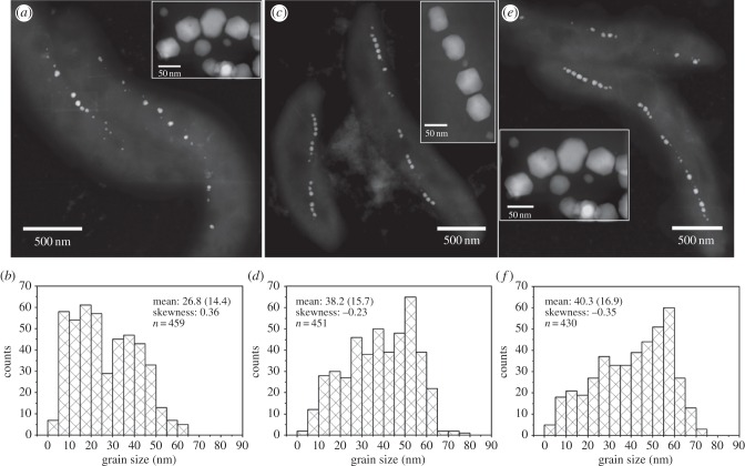
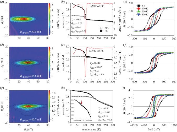
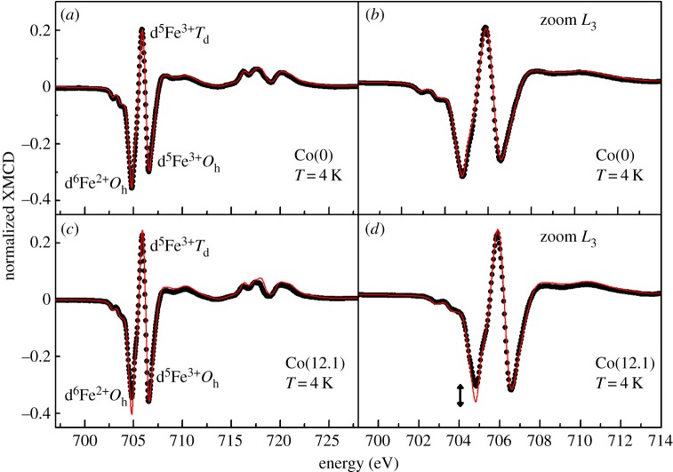
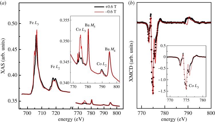
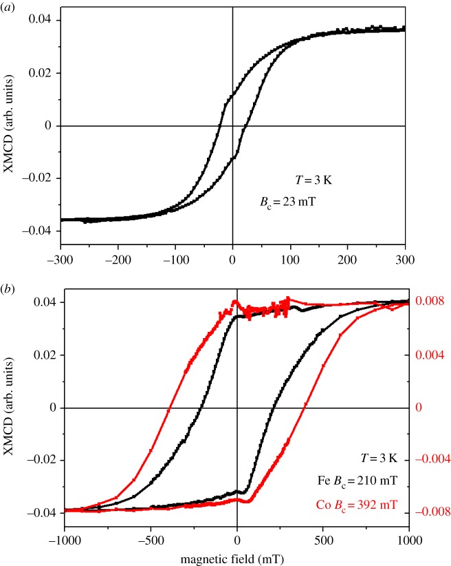
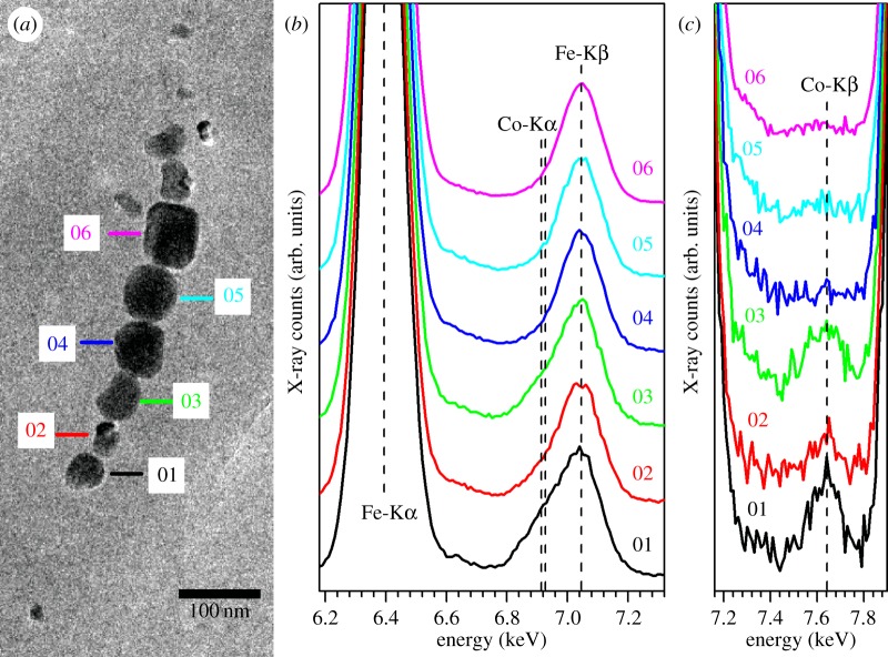
The biomineralization of magnetite nanocrystals (called magnetosomes) by magnetotactic bacteria (MTB) has attracted intense interest in biology, geology and materials science due to the precise morphology of the particles, the chain-like assembly and their unique magnetic properties. Great efforts have been recently made in producing transition metal-doped magnetosomes with modified magnetic properties for a range of applications. Despite some successful outcomes, the coordination chemistry and magnetism of such metal-doped magnetosomes still remain largely unknown. Here, we present new evidences from X-ray magnetic circular dichroism (XMCD) for element- and site-specific magnetic analyses that cobalt is incorporated in the spinel structure of the magnetosomes within Magnetospirillum magneticum AMB-1 through the replacement of Fe(2+) ions by Co(2+) ions in octahedral (Oh) sites of magnetite. Both XMCD at Fe and Co L2,3 edges, and energy-dispersive X-ray spectroscopy on transmission electron microscopy analyses reveal a heterogeneous distribution of cobalt occurring either in different particles or inside individual particles. Compared with non-doped one, cobalt-doped magnetosome sample has lower Verwey transition temperature and larger magnetic coercivity, related to the amount of doped cobalt. This study also demonstrates that the addition of trace cobalt in the growth medium can significantly improve both the cell growth and the magnetosome formation within M. magneticum AMB-1. Together with the cobalt occupancy within the spinel structure of magnetosomes, this study indicates that MTB may provide a promising biomimetic system for producing chains of metal-doped single-domain magnetite with an appropriate tuning of the magnetic properties for technological and biomedical applications.
Keywords: X-ray magnetic circular dichroism; biomineralization; cobalt-doped magnetite; coordination chemistry; magnetic alteration; magnetotactic bacteria.
© 2016 The Author(s).
Figures
Similar articles
-
Measuring spectroscopy and magnetism of extracted and intracellular magnetosomes using soft X-ray ptychography.Proc Natl Acad Sci U S A. 2016 Dec 20;113(51):E8219-E8227. doi: 10.1073/pnas.1610260114. Epub 2016 Dec 7. Proc Natl Acad Sci U S A. 2016. PMID: 27930297 Free PMC article.
-
The magnetosome membrane protein, MmsF, is a major regulator of magnetite biomineralization in Magnetospirillum magneticum AMB-1.Mol Microbiol. 2012 Aug;85(4):684-99. doi: 10.1111/j.1365-2958.2012.08132.x. Epub 2012 Jul 10. Mol Microbiol. 2012. PMID: 22716969 Free PMC article.
-
Probing the Nanostructure and Arrangement of Bacterial Magnetosomes by Small-Angle X-Ray Scattering.Appl Environ Microbiol. 2019 Nov 27;85(24):e01513-19. doi: 10.1128/AEM.01513-19. Print 2019 Dec 15. Appl Environ Microbiol. 2019. PMID: 31604767 Free PMC article.
-
Bacterial magnetosome and its potential application.Microbiol Res. 2017 Oct;203:19-28. doi: 10.1016/j.micres.2017.06.005. Epub 2017 Jun 20. Microbiol Res. 2017. PMID: 28754204 Review.
-
Applications of Magnetotactic Bacteria, Magnetosomes and Magnetosome Crystals in Biotechnology and Nanotechnology: Mini-Review.Molecules. 2018 Sep 24;23(10):2438. doi: 10.3390/molecules23102438. Molecules. 2018. PMID: 30249983 Free PMC article. Review.
Cited by
-
Single-Cell Resolution of Uncultured Magnetotactic Bacteria via Fluorescence-Coupled Electron Microscopy.Appl Environ Microbiol. 2017 May 31;83(12):e00409-17. doi: 10.1128/AEM.00409-17. Print 2017 Jun 15. Appl Environ Microbiol. 2017. PMID: 28389550 Free PMC article.
-
Therapeutic Innovations in Nanomedicine: Exploring the Potential of Magnetotactic Bacteria and Bacterial Magnetosomes.Int J Nanomedicine. 2025 Jan 11;20:403-444. doi: 10.2147/IJN.S462031. eCollection 2025. Int J Nanomedicine. 2025. PMID: 39816378 Free PMC article. Review.
-
Modifying the magnetic response of magnetotactic bacteria: incorporation of Gd and Tb ions into the magnetosome structure.Nanoscale Adv. 2022 Apr 26;4(12):2649-2659. doi: 10.1039/d2na00094f. eCollection 2022 Jun 14. Nanoscale Adv. 2022. PMID: 36132283 Free PMC article.
-
A Method for Producing Highly Pure Magnetosomes in Large Quantity for Medical Applications Using Magnetospirillum gryphiswaldense MSR-1 Magnetotactic Bacteria Amplified in Minimal Growth Media.Front Bioeng Biotechnol. 2020 Feb 18;8:16. doi: 10.3389/fbioe.2020.00016. eCollection 2020. Front Bioeng Biotechnol. 2020. PMID: 32133346 Free PMC article.
-
Renaissance for magnetotactic bacteria in astrobiology.ISME J. 2023 Oct;17(10):1526-1534. doi: 10.1038/s41396-023-01495-w. Epub 2023 Aug 17. ISME J. 2023. PMID: 37592065 Free PMC article. Review.
References
-
- Martin M, Carmona F, Cuesta R, Rondon D, Galvez N, Dominguez-Vera JM. 2014. Artificial magnetic bacteria: living magnets at room temperature. Adv. Funct. Mater. 24, 3489–3493. (10.1002/adfm.201303754) - DOI
-
- Sun S. 2006. Recent advances in chemical synthesis, self-assembly, and applications of FePt nanoparticles. Adv. Mater. 18, 393–403. (10.1002/adma.200501464) - DOI
Publication types
MeSH terms
Substances
LinkOut - more resources
Full Text Sources
Other Literature Sources
