. 2016 Sep;95(3):72.
doi: 10.1095/biolreprod.116.140905. Epub 2016 Aug 10.

Uterotonic Neuromedin U Receptor 2 and Its Ligands Are Upregulated by Inflammation in Mice and Humans, and Elicit Preterm Birth

Affiliations

Uterotonic Neuromedin U Receptor 2 and Its Ligands Are Upregulated by Inflammation in Mice and Humans, and Elicit Preterm Birth

Mathieu Nadeau-Vallée et al. Biol Reprod. 2016 Sep.

Abstract

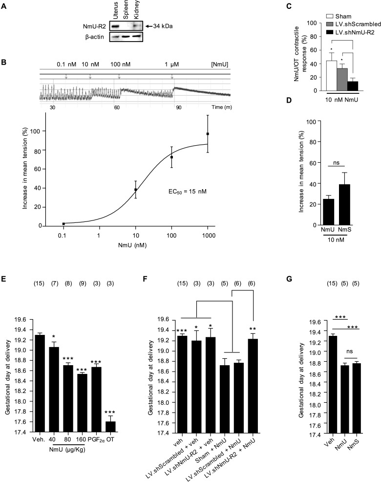
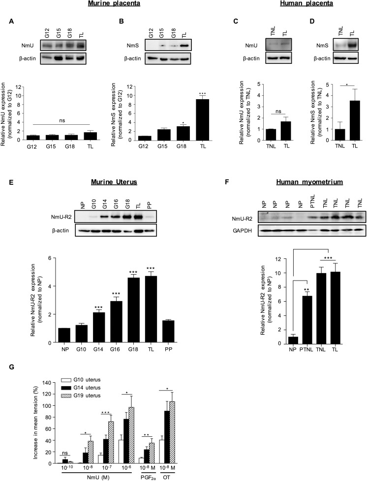
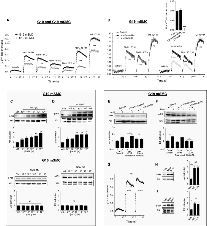
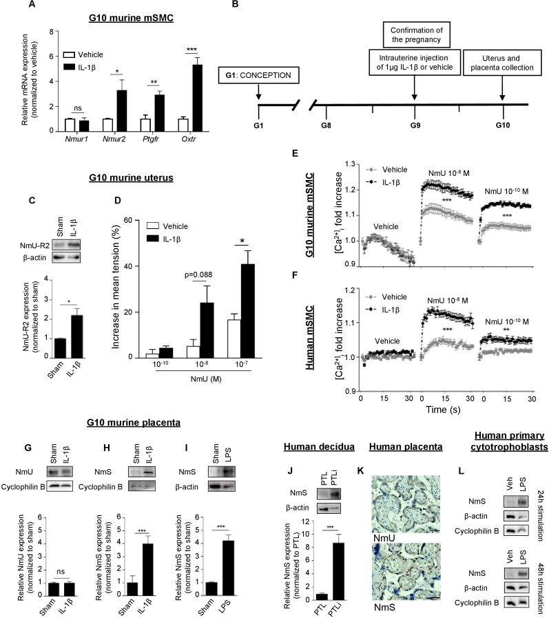
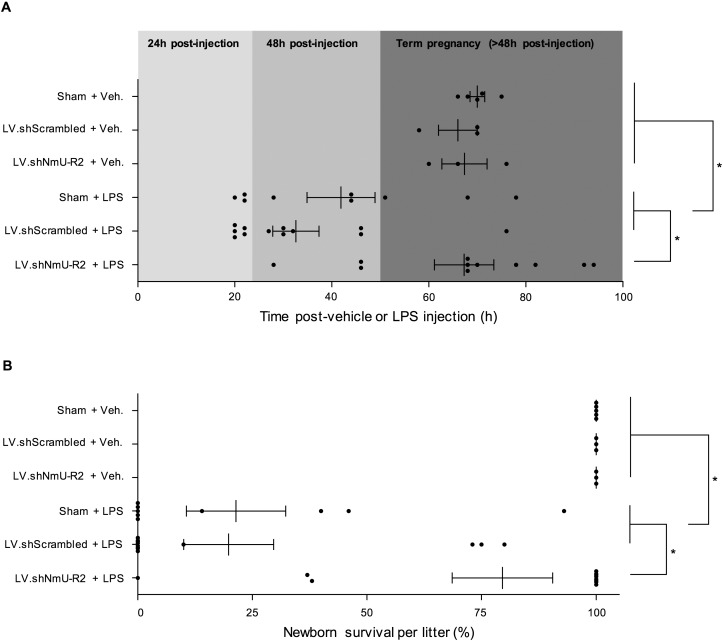
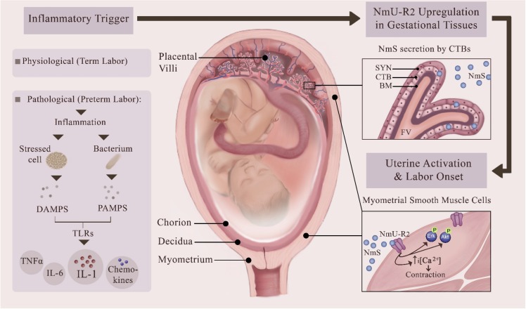
Uterine labor requires the conversion of a quiescent (propregnancy) uterus into an activated (prolabor) uterus, with increased sensitivity to endogenous uterotonic molecules. This activation is induced by stressors, particularly inflammation in term and preterm labor. Neuromedin U (NmU) is a neuropeptide known for its uterocontractile effects in rodents. The objective of the study was to assess the expression and function of neuromedin U receptor 2 (NmU-R2) and its ligands NmU and the more potent neuromedin S (NmS) in gestational tissues, and the possible implication of inflammatory stressors in triggering this system. Our data show that NmU and NmS are uterotonic ex vivo in murine tissue, and they dose-dependently trigger labor by acting specifically via NmU-R2. Expression of NmU-R2, NmU, and NmS is detected in murine and human gestational tissues by immunoblot, and the expression of NmS in placenta and of NmU-R2 in uterus increases considerably with gestation age and labor, which is associated with amplified NmU-induced uterocontractile response in mice. NmU- and NmS-induced contraction is associated with increased NmU-R2-coupled Ca++ transients, and Akt and Erk activation in murine primary myometrial smooth muscle cells (mSMCs), which are potentiated with gestational age. NmU-R2 is upregulated in vitro in mSMCs and in vivo in uterus in response to proinflammatory interleukin 1beta (IL1beta), which is associated with increased NmU-induced uterocontractile response and Ca++ transients in murine and human mSMCs; additionally, placental NmS is markedly upregulated in vivo in response to IL1beta. In human placenta at term, immunohistological analysis revealed NmS expression primarily in cytotrophoblasts; furthermore, stimulation with lipopolysaccharide (LPS; Gram-negative endotoxin) markedly upregulates NmS expression in primary human cytotrophoblasts isolated from term placentas. Correspondingly, decidua of women with clinical signs of infection who delivered preterm display significantly higher expression of NmS compared with those without infection. Importantly, in vivo knockdown of NmU-R2 prevents LPS-triggered preterm birth in mice and the associated neonatal mortality. Altogether, our data suggest a critical role for NmU-R2 and its ligands NmU and NmS in preterm labor triggered by infection. We hereby identify NmU-R2 as a relevant target for preterm birth.

Keywords: NmU-R2; calcium; contraction; infection; inflammation; neuromedin S; neuromedin U; preterm birth; preterm labor; uterine labor.


Figures

FIG. 1
FIG. 1
NmU and NmS induce uterine contractions and labor via NmU-R2. A) NmU-R2 immunoblot from term uterus. Spleen and kidney samples were used as negative and positive controls, respectively. B) Representative ex vivo contraction assay performed on a myometrium fragment from a pregnant mouse at term in response to increasing NmU concentrations (top) and dose-response curve (bottom). C) Pregnant mice were pretreated with LV.shNmU-R2 or LV.shScrambled at G15, and their uteri were collected at term for a contraction assay in response to NmU and OT. For each uterine strip, the contractile response to NmU was normalized to its response to OT (to control for interindividual variability). *P < 0.05. D) Ex vivo contraction assay on G19 uteri in response to 10−8 M NmU or NmS. E) Pregnant mice were injected intraperitoneally with increasing NmU doses twice a day from G17 to G18. Control animals were given an equivalent volume of saline. PGF and OT were used as positive controls at a dose of 160 μg/kg. *P < 0.05, ***P < 0.001 compared with vehicle only. F) Pregnant mice were pretreated with LV.shNmU-R2 or LV.shScrambled at G15 and then treated with 160 μg/kg NmU twice a day from G17 to G18. *P < 0.05, **P < 0.01, ***P < 0.001 compared with NmU or NmU + LV.shScrambled. G) Pregnant mice were injected intraperitoneally with 160 μg/kg NmS or NmU twice a day from G17 to G18. ***P < 0.001 compared with vehicle only. Data for AD are representative of four to five mice per group. The number of mice used in EG is displayed above each group, and mice treated with vehicle (control mice) were pooled together and repeatedly shown in each graph. Values are presented as mean ± SEM. Data were statistically analyzed using one-way ANOVA with comparison to control groups using Dunnett multiple-comparison test; ns, not significant.
FIG. 2
FIG. 2
The expression of NmS in placenta and of NmU-R2 in uterus increases near term and during labor in mice and humans, which is associated with increased NmU-induced myometrial contractility at term. A and B) NmU (A) and NmS (B) representative immunoblots of murine placentas collected at different gestational ages (G) and during spontaneous term labor (TL). Lower panels show densitometric analysis of protein bands normalized with β-actin and plotted as fold change versus the control group (G12). C and D) NmU (C) and NmS (D) immunoblots of human placenta biopsies from women at term not in labor (TNL) or in established labor (TL). Lower panels show densitometric analysis of protein bands normalized with β-actin and plotted as fold change versus the control group (TNL) of all patients screened (TNL, n = 6; TL, n = 6). E) NmU-R2 representative immunoblot of murine uteri collected at different gestational ages (G), during spontaneous term labor (TL) and 24 h postpartum (PP). The lower panel shows densitometric analysis of protein bands normalized with β-actin and plotted as fold change versus the control group (NP). F) Representative immunoblot of NmU-R2 from human myometrial tissue biopsies from four nonpregnant (NP) women (hysterectomy), one pregnant women at preterm without any clinical sign of labor (PTNL), and four pregnant women at term without any clinical sign of labor (TNL). The lower panel shows densitometric analysis of protein bands normalized with β-actin and plotted as fold change versus the control group (NP) of all patients screened (NP, n = 4; PTNL, n = 4; TNL, n = 9; TL, n = 5), as presented in F and Supplemental Figure S2G. G) Ex vivo contraction assay in response to increasing doses of NmU performed on myometrium fragments from pregnant mice at G10, G14, or G19. Uteri collected at G19 were only considered if the mice were still undelivered. PGF and OT were used as positive controls at a dose of 10−8 M. Data are representative of four to five mice per group and at least three independent experiments. Values are presented as mean ± SEM. *P < 0.05, **P < 0.01, ***P < 0.001 by one-way ANOVA with comparison to control groups using Dunnett multiple-comparison test; ns, not significant.
FIG. 3
FIG. 3
NmU-R2-associated signaling in mSMCs is potentiated by gestational age. A) Calcium assay performed on primary mSMCs from pregnant mice at either G10 or G19. PGF and OT were used as positive controls at a concentration of 10−6 M; n = 12–45 in each group. B) G19 myometrial cells were pretreated with LV.shNmU-R2 or LV.shScrambled for 72 h and then used in a calcium assay in response to 10−8 M NmU or OT. The calcium mobilization response to NmU is also presented when normalized with the response to OT (inset); n = 6–12 in each group. C and D) Akt (C) and Erk (D) phosphorylation immunoreactivity on primary mSMCs from pregnant mice at G10 or G19 stimulated with increasing concentrations of NmU for 10 min. Data are representative of four to five independent experiments. E and F) Akt (E) and Erk (F) immunoreactivity on G19 myometrial cells pretreated with LV.shNmU-R2 or LV.shSrambled for 72 h and then stimulated with 10−6 M NmU for 10 min. Data are representative of four to five independent experiments. G) Calcium assay of G19 myometrial cells treated with 10−6 M NmU or NmS; n = 6–45 in each group. H and I) Akt (H) and Erk (I) phosphorylation immunoblot of G19 myometrial cells treated with 10−6 M NmU or NmS for 10 min. Data are representative of four to five independent experiments. *P < 0.05, **P < 0.01, ***P < 0.001 by one-way ANOVA with comparison to control groups using Dunnett multiple-comparison test; ns, not significant.
FIG. 4
FIG. 4
NmU-R2 in uterus and NmS in placenta are under the control of inflammation in mice and humans. A) Quantitative PCR performed on primary mSMCs collected from pregnant mice at G10 and stimulated with 5 ng/ml IL1β for 6 h. Results are relative to 18S and plotted as fold over the control group (vehicle). B) Schematic representation of the in vivo model used. C) Representative NmU-R2 immunoblot (top) and densitometric analysis (bottom) of uteri of pregnant mice collected 24 h after an intrauterine injection of saline (sham) or IL1β. D) Ex vivo contraction assay performed on uteri from pregnant mice exposed for 24 h to saline (sham) or IL1β. Data are representative of three to four mice per group. E and F) Pregnant mouse (G10) mSMCs or human mSMCs (hTERT-C3 cell line) were treated for 24 h with IL1β or vehicle, and were used in a calcium assay; n = 6–12 in each group. G and H) Immunoblots (top) and relative densitometric analysis of protein bands (bottom) showing NmU (G) and NmS (H) expression in placenta of pregnant mice 24 h after an intrauterine injection of either saline (sham) or IL1β. I) Immunoblot (top) and relative densitometric analysis of protein bands (bottom) showing NmS expression in placenta of pregnant mice 24 h after an intrauterine injection of either saline (sham) or LPS. J) NmS immunoblot from human decidual biopsies from women in preterm labor with (PTLi, n = 3) and without (PTL, n = 3) clinical evidence of infection. Bottom panel shows densitometric analysis of protein bands normalized with β-actin and plotted as fold over the control group (PTL). K) Immunohistochemistry analysis performed on term human placentas blotted against NmU and NmS. For the NmU panel, black arrows represent syncytiotrophoblasts; for the NmS panel, black arrows represent cytotrophoblasts and white arrows Hofbauer cells. Bar = 40 μm. L) Primary human cytotrophoblasts were stimulated with LPS for 24 h (top) or 48 h (bottom), and cell lysates were blotted against NmS. β-Actin and cyclophilin B were used as loading controls. Values are presented as mean ± SEM. Data are representative of at least three independent experiments. *P < 0.05, **P < 0.01, ***P < 0.001 by one-way ANOVA with comparison to control groups using Dunnett multiple-comparison test; ns, not significant.
FIG. 5
FIG. 5
NmU-R2 is important for infection-induced adverse gestation outcomes. A and B) Pregnant mice were pretreated with intrauterine injections of vehicle (sham), LV.shScrambled, or LV.shNmU-R2 at G13 (one injection in each uterine horn), and then treated with a single intraperitoneal injection of 10 μg of LPS or an equivalent volume of saline at G16. The timing of birth (A) and the survival of pups at birth per litter (B) were rigorously assessed. Values are presented as mean ± SEM. *P < 0.05 by one-way ANOVA with comparison to control groups using Dunnett multiple-comparison test.
FIG. 6
FIG. 6
Proposed role of NmU-R2 and its ligand NmS in physiological term labor and in pathological PTB. The schema provides a current view of inflammation-associated preterm birth whereby pathogen-associated molecular patterns (PAMPs) and damage-associated molecular patterns (DAMPs) activate pattern recognition receptors in uterus, predominantly Toll-like receptors (TLRs), to trigger an inflammatory cascade characterized by the local maturation and release of proinflammatory, prolabor cytokines, leading to uterine activation and PTB. Our study reveals that uterotonic NmS and its cognate receptor NmU-R2 are endogenously produced in human placenta and myometrium, respectively, and are upregulated by the PAMP LPS and by its downstream mediator IL1. We propose that this neuromedin system is implicated in sustaining or initiating uterine contractions in both physiological labor and pathological preterm labor associated with inflammation. This figure was made exclusively for this manuscript by the authors.

Similar articles

Cited by

References

    1. Cook JL, Shallow MC, Zaragoza DB, Anderson KI, Olson DM. Mouse placental prostaglandins are associated with uterine activation and the timing of birth. Biol Reprod. 2003;68:579–587. - PubMed
    1. Romero R, Espinoza J, Gonçalves LF, Kusanovic JP, Friel LA, Nien JK. Inflammation in preterm and term labour and delivery. Semin Fetal Neonatal Med. 2006;11:317–326. - PMC - PubMed
    1. Bennett P. Preterm labour. In: Edmonds DK, editor. Dewhurst's Textbook of Obstetrics and Gynaecology, 8th ed. Oxford, UK: Wiley-Blackwell; 2012. pp. 338–355. In: (ed.)
    1. Christiaens I, Zaragoza DB, Guilbert L, Robertson SA, Mitchell BF, Olson DM. Inflammatory processes in preterm and term parturition. J Reprod Immunol. 2008;79:50–57. - PubMed
    1. Keelan JA, Blumenstein M, Helliwell RJ, Sato TA, Marvin KW, Mitchell MD. Cytokines, prostaglandins and parturition–a review. Placenta. 2003;24(suppl A):S33–S46. - PubMed