Review
. 2016 Aug 3:8:25-36.
doi: 10.4137/GEG.S31863. eCollection 2016.

The Evolution of Epigenetics: From Prokaryotes to Humans and Its Biological Consequences

Affiliations
Review

The Evolution of Epigenetics: From Prokaryotes to Humans and Its Biological Consequences

Amber Willbanks et al. Genet Epigenet. .

Abstract

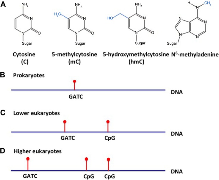
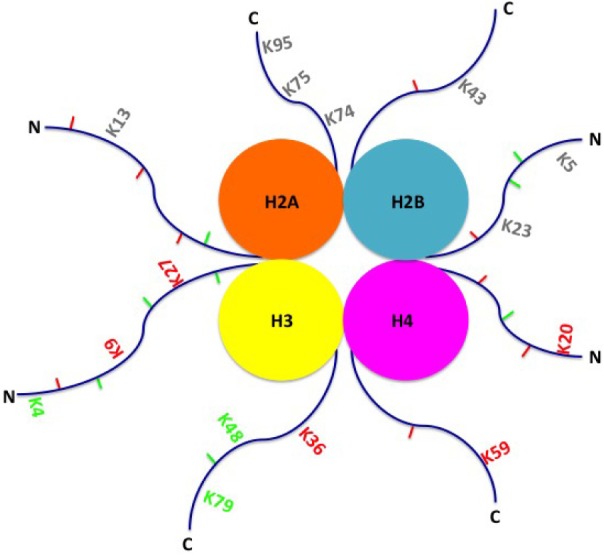
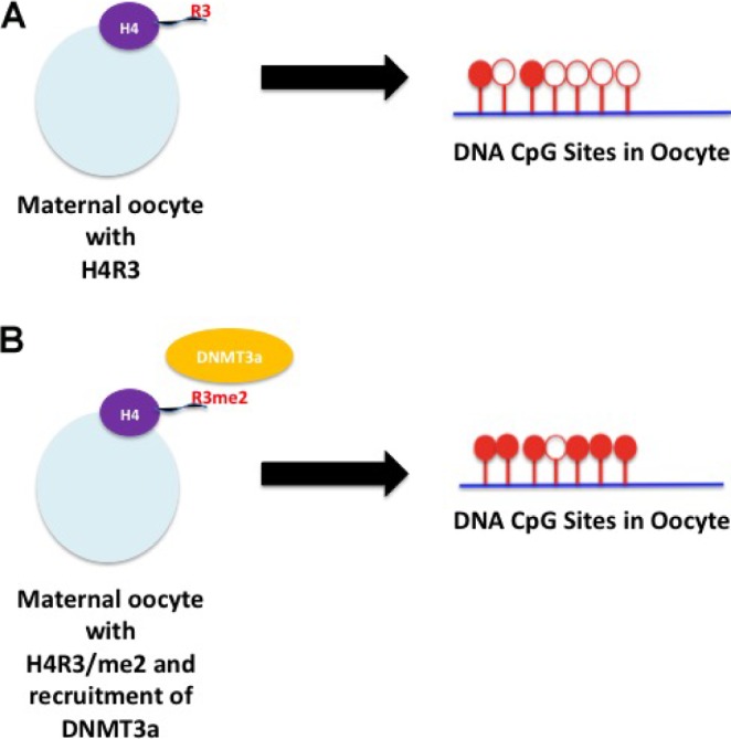
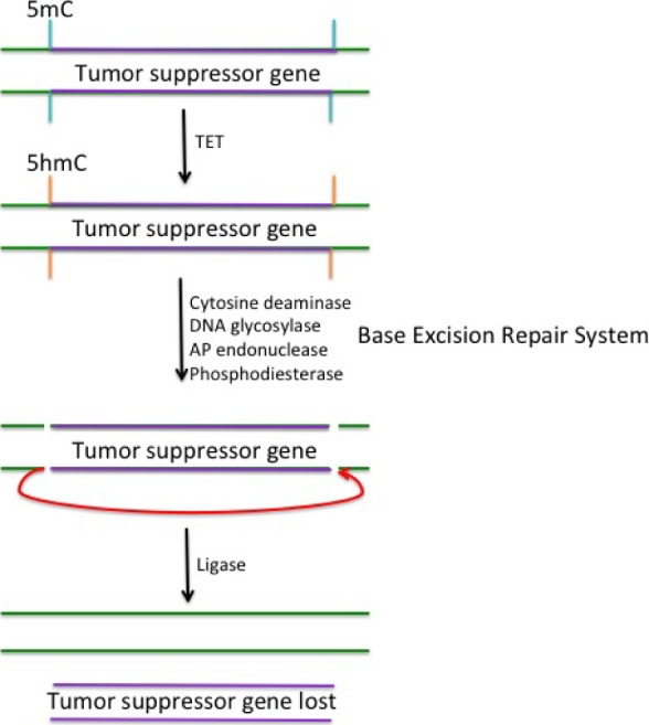
The evolution process includes genetic alterations that started with prokaryotes and now continues in humans. A distinct difference between prokaryotic chromosomes and eukaryotic chromosomes involves histones. As evolution progressed, genetic alterations accumulated and a mechanism for gene selection developed. It was as if nature was experimenting to optimally utilize the gene pool without changing individual gene sequences. This mechanism is called epigenetics, as it is above the genome. Curiously, the mechanism of epigenetic regulation in prokaryotes is strikingly different from that in eukaryotes, mainly higher eukaryotes, like mammals. In fact, epigenetics plays a significant role in the conserved process of embryogenesis and human development. Malfunction of epigenetic regulation results in many types of undesirable effects, including cardiovascular disease, metabolic disorders, autoimmune diseases, and cancer. This review provides a comparative analysis and new insights into these aspects.

Keywords: diseases; epigenetics; eukaryotes; evolution; mammals; prokaryotes.


Figures

Figure 1
Figure 1
Differential DNA methylation patterns in prokaryotes, lower eukaryotes, and higher eukaryotes. (A) Pictorial representation of cytosine, 5-methylcytosine, 5-hydroxymethylcytosine, and N6-methyladenine; (B) prokaryote, (C) lower eukaryote, and (D) higher eukaryote DNA methylation is denoted in red on either adenine (A) or cytosine (C).
Figure 2
Figure 2
Activating and inhibitory histone modifications. Histone modifications include methylation and demethylation on lysine and arginine residues, ubiquitination, and sumoylation. Activating modifications (green), inhibitory modifications (red), and modifications with unknown function (gray) are shown on either the C-terminus (C) or N-terminus (N) of histone tails. Lysine methylations are marked with K and the number of the residue methylated.
Figure 3
Figure 3
Differential methylation of H4R in the maternal oocyte regulates the methylation status of DNA during embryogenesis. (A) No maternal oocyte H4R3 methylation yields little DNA CpG site methylation. (B) Maternal oocyte H4R3 methylation yields more methylation of DNA CpG sites. Red indicates methylation and empty circles represent nonmethylation. DNMT3a denotes DNA methyltransferase 3a.
Figure 4
Figure 4
Gene deletion due to base excision of hydroxymethylcytosine. Demethylation of 5-methylctyosine (5mC) is catalyzed by the TET enzymes. TET converts 5-methylcytosine (indicated in blue) to 5-hydroxymethylcytosine (5hmC; indicated in orange). 5hmC may subsequently be acted upon by cytosine deaminase enzymes, resulting in a base mismatch that can be targeted by the base excision repair system to correct the mismatch. DNA glycosylase enzymes act to remove the base, creating an AP site. AP endonuclease and phosphodiesterase enzymes act on the AP site to excise the phosphate backbone where the 5mC used to be, creating a lesion in the DNA to be filled in by DNA polymerase. However, if both ends of the gene have bases excised at the same time and on both strands, it is possible for the DNA to religate, bringing the opposite ends of the gene together, and removing the gene from the genome. This model depicts a tumor suppressor gene (indicated in purple) being lost from the rest of the DNA (green).

References

    1. Turner BM. Epigenetic responses to environmental change and their evolutionary implications. Philos Trans R Soc Lond B. 2009;364:1534. - PMC - PubMed
    1. Eid A, Zawia N. Consequences of lead exposure, and its emerging role as an epigenetic modifier in the aging brain. Neurotoxicology. 2016 doi: 10.1016/j.neuro.2016.04.006. - DOI - PubMed
    1. Casey SC, Vaccari M, Al-Mulla F, et al. The effect of environmental chemicals on the tumor microenvironment. Carcinogenesis. 2015;36:S160–S183. - PMC - PubMed
    1. Chai H, Brown RE. Field effect in cancer: an update. Ann Clin Lab Sci. 2009;39(4):331–337. - PubMed
    1. Bollati V, Baccarelli A, Hou L, et al. Changes in DNA methylation patterns in subjects exposed to low-dose benzene. Cancer Res. 2007;67:876–880. - PubMed

LinkOut - more resources