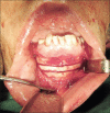
Anterior Palatal Island Advancement Flap for Bone Graft Coverage: Technical Note
- PMID: 27512552
- PMCID: PMC4966204
- DOI: 10.4103/2006-8808.185655
Anterior Palatal Island Advancement Flap for Bone Graft Coverage: Technical Note
Abstract
Background: The most important step in bone graft management is soft tissue coverage. Dehiscence of the wound leads to graft exposure and subsequent problems.
Purpose: This study introduces an axial pattern flap for bone graft coverage in anterior maxilla.
Patients and methods: Use of Anterior Palatal Island Advancement Flap is presented by the authors. It is a mucoperiosteal flap with axial pattern blood supply, based on nasopalatine artery. It is easy to raise and predictable.
Results: Anterior Palatal Island Advancement Flap was effective in bone graft coverage in premaxillary edentulous area.
Conclusion: It can be used as an aid for bone graft coverage of premaxillary edentulous ridge, where the need for mucosa is small in width but long in length.
Keywords: Anterior maxilla; bone graft; dental implant; palatal advancement flap.
Figures


Similar articles
-
Nasopalatine Island Advancement Flap.J Craniofac Surg. 2016 Nov;27(8):2190-2191. doi: 10.1097/SCS.0000000000003094. J Craniofac Surg. 2016. PMID: 28005787
-
Vascularized connective tissue flap for bone graft coverage.J Oral Implantol. 2011 Apr;37(2):279-85. doi: 10.1563/AAID-JOI-D-09-00146.1. Epub 2010 Jun 16. J Oral Implantol. 2011. PMID: 20553154
-
Relationship of central incisor implant placement to the ridge configuration anterior to the nasopalatine canal in dentate and partially edentulous individuals: a comparative study.PeerJ. 2015 Nov 3;3:e1315. doi: 10.7717/peerj.1315. eCollection 2015. PeerJ. 2015. PMID: 26557434 Free PMC article.
-
Nasoplatine Island Advancement Flap for Closure of Anterior Palatal Fistula in Patient With Isolated Cleft Palate.J Craniofac Surg. 2019 Jul;30(5):e462-e463. doi: 10.1097/SCS.0000000000005549. J Craniofac Surg. 2019. PMID: 31299815
-
A bilateral pediculated palatal periosteal connective tissue flap for coverage of large bone grafts in the anterior maxillary region.Iran J Otorhinolaryngol. 2012 Summer;24(68):143-6. Iran J Otorhinolaryngol. 2012. PMID: 24303400 Free PMC article.
References
-
- Chaushu G, Mardinger O, Peleg M, Ghelfan O, Nissan J. Analysis of complications following augmentation with cancellous block allografts. J Periodontol. 2010;81:1759–64. - PubMed
-
- Garg AK. Subnasal elevation and bone augmentation in dental implantology. Dent Implantol Update. 2008;19:17–21. - PubMed
-
- Heller AL, Heller RL, Cook G, D’Orazio R, Rutkowski J. Soft tissue management techniques for implant dentistry: A clinical guide. J Oral Implantol. 2000;26:91–103. - PubMed
-
- Abrahamsson P, Wälivaara DÅ, Isaksson S, Andersson G. Periosteal expansion before local bone reconstruction using a new technique for measuring soft tissue profile stability: A clinical study. J Oral Maxillofac Surg. 2012;70:e521–30. - PubMed
-
- Nemcovsky CE, Moses O, Artzi Z, Gelernter I. Clinical coverage of dehiscence defects in immediate implant procedures: Three surgical modalities to achieve primary soft tissue closure. Int J Oral Maxillofac Implants. 2000;15:843–52. - PubMed
LinkOut - more resources
Full Text Sources
Other Literature Sources

