

Adult medulloblastoma: A rare case report and literature review
- PMID: 27512610
- PMCID: PMC4960923
- DOI: 10.4103/2152-7806.185782
Adult medulloblastoma: A rare case report and literature review
Abstract
Background: Medulloblastoma is a highly malignant embryonal tumor which commonly arises in the cerebellum. It is relatively rare and accounts for less than 2% of all primary brain tumors. The tumor primarily occurs in childhood; however, rarely, it may be found in adult population. In addition, medulloblastoma in adult population shows features which are quite distinct from the pediatric group.
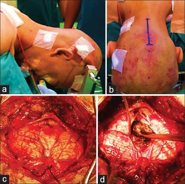
Case description: We report the case of a 33-year-old man who presented to our institution with a history of blurred vision of both eyes for 5 months preceded by intermittent headache since the previous year. Preoperative investigation suggested a posterior fossa mass and we suspected an ependymoma. The patient underwent ventriculoperitoneal shunt and craniotomy tumor removal, followed by radiotherapy. Histopathological and immunohistochemical examination were performed, and the results showed a diagnosis of medulloblastoma.
Conclusion: This case is exceptional because adult medulloblastoma occurrence in our center is extremely rare, and the diagnosis can only be established through histopathological and immunohistochemical studies.
Keywords: A rare case of adult medulloblastoma; fossa posterior brain tumor; immunohistochemical studies.
Figures


References
-
- Association ABT. Chicago: 2012. American Brain Tumor Association. Medulloblastoma.
-
- Bourgouin PM, Tampieri D, Grahovac SZ, Léger C, Carpio RD, Melançon D. CT and MR imaging findings in adults with cerebellar medulloblastoma: Comparison with findings in children. Am J Roentgenol. 1992;159:609–12. - PubMed
-
- Brandes AA, Franceschi E, Tosoni A, Reni M, Gatta G, Vecht C, et al. Adult neuroectodermal tumors of posterior fossa (medulloblastoma) and of supratentorial sites (stPNET) Crit Rev Oncol Hematol. 2009;71:165–79. - PubMed
-
- Brandes AA, Paris MK. Review of the prognostic factors in medulloblastoma of children and adults. Crit Rev Oncol Hematol. 2004;50:121–8. - PubMed
Publication types
LinkOut - more resources
Full Text Sources
Other Literature Sources