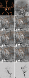
CBCT-based 3D MRA and angiographic image fusion and MRA image navigation for neuro interventions
- PMID: 27512846
- PMCID: PMC4985301
- DOI: 10.1097/MD.0000000000004358
CBCT-based 3D MRA and angiographic image fusion and MRA image navigation for neuro interventions
Abstract
Digital subtracted angiography (DSA) remains the gold standard for diagnosis of cerebral vascular diseases and provides intraprocedural guidance. This practice involves extensive usage of x-ray and iodinated contrast medium, which can induce side effects. In this study, we examined the accuracy of 3-dimensional (3D) registration of magnetic resonance angiography (MRA) and DSA imaging for cerebral vessels, and tested the feasibility of using preprocedural MRA for real-time guidance during endovascular procedures.Twenty-three patients with suspected intracranial arterial lesions were enrolled. The contrast medium-enhanced 3D DSA of target vessels were acquired in 19 patients during endovascular procedures, and the images were registered with preprocedural MRA for fusion accuracy evaluation. Low-dose noncontrasted 3D angiography of the skull was performed in the other 4 patients, and registered with the MRA. The MRA was overlaid afterwards with 2D live fluoroscopy to guide endovascular procedures.The 3D registration of the MRA and angiography demonstrated a high accuracy for vessel lesion visualization in all 19 patients examined. Moreover, MRA of the intracranial vessels, registered to the noncontrasted 3D angiography in the 4 patients, provided real-time 3D roadmap to successfully guide the endovascular procedures. Radiation dose to patients and contrast medium usage were shown to be significantly reduced.Three-dimensional MRA and angiography fusion can accurately generate cerebral vasculature images to guide endovascular procedures. The use of the fusion technology could enhance clinical workflow while minimizing contrast medium usage and radiation dose, and hence lowering procedure risks and increasing treatment safety.
Conflict of interest statement
The author(s) declared no potential conflicts of interest with respect to the research, authorship, and/or publication of this article.
Figures


Similar articles
-
Feasibility of three-dimensional magnetic resonance angiography-fluoroscopy image fusion technique in guiding complex endovascular aortic procedures in patients with renal insufficiency.J Vasc Surg. 2017 May;65(5):1440-1452. doi: 10.1016/j.jvs.2016.10.083. Epub 2016 Dec 23. J Vasc Surg. 2017. PMID: 28017584
-
Three-dimensional image fusion of CTA and angiography for real-time guidance during neurointerventional procedures.J Neurointerv Surg. 2017 Mar;9(3):302-306. doi: 10.1136/neurintsurg-2015-012216. Epub 2016 Apr 5. J Neurointerv Surg. 2017. PMID: 27048959
-
Follow-up of intracranial aneurysms treated by flow diverter: comparison of three-dimensional time-of-flight MR angiography (3D-TOF-MRA) and contrast-enhanced MR angiography (CE-MRA) sequences with digital subtraction angiography as the gold standard.J Neurointerv Surg. 2016 Jan;8(1):81-6. doi: 10.1136/neurintsurg-2014-011449. Epub 2014 Oct 28. J Neurointerv Surg. 2016. PMID: 25352582
-
MRA versus DSA for the follow-up imaging of intracranial aneurysms treated using endovascular techniques: a meta-analysis.J Neurointerv Surg. 2019 Oct;11(10):1009-1014. doi: 10.1136/neurintsurg-2019-014936. Epub 2019 May 2. J Neurointerv Surg. 2019. PMID: 31048457 Review.
-
Usefulness of Different Imaging Methods in the Diagnosis of Cerebral Vasculopathy.Neuroimaging Clin N Am. 2024 Feb;34(1):39-52. doi: 10.1016/j.nic.2023.07.001. Epub 2023 Aug 8. Neuroimaging Clin N Am. 2024. PMID: 37951704 Review.
Cited by
-
The Usefulness of a 3D Roadmap of Occluded Vessels Created from Rapid 3D Proton Density-Weighted Imaging for Mechanical Thrombectomy.J Neuroendovasc Ther. 2025;19(1):2024-0044. doi: 10.5797/jnet.tn.2024-0044. Epub 2024 Nov 6. J Neuroendovasc Ther. 2025. PMID: 40018276 Free PMC article.
-
Computed tomography cavernosography combined with volume rendering to observe venous leakage in young patients with erectile dysfunction.Br J Radiol. 2018 Nov;91(1091):20180118. doi: 10.1259/bjr.20180118. Epub 2018 Aug 13. Br J Radiol. 2018. PMID: 30028186 Free PMC article.
-
Using temporal and structural data to reconstruct 3D cerebral vasculature from a pair of 2D digital subtraction angiography sequences.Comput Med Imaging Graph. 2022 Jul;99:102076. doi: 10.1016/j.compmedimag.2022.102076. Epub 2022 May 21. Comput Med Imaging Graph. 2022. PMID: 35636377 Free PMC article.
-
Simulation of superselective catheterization for cerebrovascular lesions using a virtual injection software.CVIR Endovasc. 2021 Jun 14;4(1):52. doi: 10.1186/s42155-021-00242-6. CVIR Endovasc. 2021. PMID: 34125300 Free PMC article.
-
Bimodal ERCP, a new way of seeing things.Endosc Int Open. 2020 Mar;8(3):E368-E376. doi: 10.1055/a-1070-8749. Epub 2020 Feb 21. Endosc Int Open. 2020. PMID: 32118109 Free PMC article.
References
-
- Siewerdsen JH, Moseley D, Burch S, et al. Volume CT with a flat-panel detector on a mobile, isocentric C-arm: pre-clinical investigation in guidance of minimally invasive surgery. Med Phys 2005; 32:241–254. - PubMed
-
- Benndorf G, Klucznik RP, Strother CM. Images in cardiovascular medicine. Angiographic computed tomography for imaging of under deployed intracranial stent. Circulation 2006; 114:e499–e500. - PubMed
-
- Akpek S, Brunner T, Benndorf G, et al. Three-dimensional imaging and cone beam volume CT in C-arm angiography with flat panel detector. Diagn Interv Radiol 2005; 11:10–13. - PubMed
Publication types
MeSH terms
LinkOut - more resources
Full Text Sources
Other Literature Sources
Medical

