

Review
doi: 10.1016/j.chom.2016.07.010.
Discrimination and Integration of Stress Signals by Pathogenic Bacteria
Affiliations
- PMID: 27512902
- PMCID: PMC5111874
- DOI: 10.1016/j.chom.2016.07.010
Item in Clipboard
Review
Discrimination and Integration of Stress Signals by Pathogenic Bacteria
Cell Host Microbe.
.
Abstract
For pathogenic bacteria, the ability to sense and respond to environmental stresses encountered within the host is critically important, allowing them to adapt to changing conditions and express virulence genes appropriately. This review considers the diverse molecular mechanisms by which stress conditions are sensed by bacteria, how related signals are discriminated, and how stress responses are integrated, highlighting recent studies in selected bacterial pathogens of clinical relevance.
Published by Elsevier Inc.
Figures
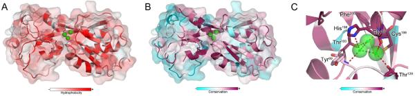
The Pseudomonas aeruginosa OxyR structure suggests a role for H2O molecules in H2O2 sensing. (A) Peroxidatic Cys199 is in a hydrophobic pocket of OxyR. Hydrophobic and hydrophilic residues are shown in red and white, respectively. Panel B shows the structure of PaOxyR with the conserved residues forming the pocket where the three H2O molecules reside. Conserved amino acids (purple) were identified using the ConSurf server with input of 75 OxyR orthologs identified by PSI-BLAST in the SWISS-PROT protein sequence database (Celniker et al., 2013; Ashkenazy et al., 2010). (C) Three H2O molecules (green spheres) establish a network of hydrogen bonding with amino acid residues in the vicinity of Cys199. Red dashes show putative hydrogen-bonding of H2O with the backbone, and side-chains of conserved residues. The H2O molecule most proximal to Cys199 is replaced by H2O2. The two H2O molecules remaining in the pocket aid in the protonation of a leaving OH− group arising from peroxidatic cleavage of H2O2 by the Cys199-reduced thiol. Residues interacting with the three H2O molecules are highly conserved (purple) in OxyR, suggesting a common mechanism of H2O2 sensing among OxyR orthologs. PDB code 4Y0M was used for PyMol analysis.
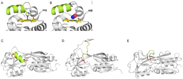
(A) Reduced H-NOX from Shewanella oneidensis with the penta-coordinated heme (yellow) bound to His103. (B) Binding of NO· (blue and red spheres) to the H-NOX heme severs the bond between His103 and Fe2+. Free His103 rotates the αF helix (green), activating signal transduction. Recent structures of the H2O2 sensor OxyR from Pseudomonas aeruginosa have revealed the molecular mechanism of sensing and signal transduction in response to H2O2. (C) Peroxidatic Cys199 and Cys208 are 15–17Å apart in opposite ends of an α-helix (green). (D) Oxidation of Cys199 by H2O2 produces a sulfenic acid (-SOH) intermediate that is mimicked by a C199D mutation. Oxidation of Cys199 triggers new hydrogen-bonding interactions between the sulfenylated group and Arg201 and Phe200 (not shown). (E) The new hydrogen-bonding disorganizes the intervening α-helix, increasing the reactivity of Cys208 and facilitating formation of a disulfide. (C) and (D) show structures of reduced and OxyR C199D proteins from P. aeruginosa, whereas (E) shows oxidized OxyR from Escherichia coli. PDB codes 4U99, 4U9B, 4Y0M, 4XWS, and 1IBA were used to generate the images in panels (A–E), respectively.

Dedicated sensors of reactive species discriminate closely related diatomic gases. H-NOX proteins from Caldoanaerobacter subterraneus (A) and Shewanella oneidensis (B) can discriminate between O2 and NO·, respectively. The heme (yellow) of C. subterraneus H-NOX is hexa-coordinated with O2, the porphyrin ring, and the imidazole group of His102. O2 maintains hydrogen-bonding interactions (red dashes) with the hydroxyl group of Tyr140. In contrast, the heme of NO·-dedicated sensors such as S. oneidensis H-NOX is penta-coordinated. Panel (B) shows the NO· molecule (red and blue spheres) in the proximal face of the porphyrin ring. The short side-chain of Cys141 does not allow hydrogen-bonding after NO· binding at the distal face of the porphyrin ring (not shown). PDB codes 1U55 and 4U9B were used for the analysis of C. subterraneus H-NOX and S. oneidensis H-NOX, respectively.
References
Publication types
MeSH terms
Substances
Grants and funding
LinkOut - more resources
Full Text Sources
Other Literature Sources