. 2016 Aug 11;12(8):e1005876.
doi: 10.1371/journal.pgen.1005876. eCollection 2016 Aug.

Combating a Global Threat to a Clonal Crop: Banana Black Sigatoka Pathogen Pseudocercospora fijiensis (Synonym Mycosphaerella fijiensis) Genomes Reveal Clues for Disease Control

Affiliations

Combating a Global Threat to a Clonal Crop: Banana Black Sigatoka Pathogen Pseudocercospora fijiensis (Synonym Mycosphaerella fijiensis) Genomes Reveal Clues for Disease Control

Rafael E Arango Isaza et al. PLoS Genet. .

Erratum in

Abstract

Black Sigatoka or black leaf streak disease, caused by the Dothideomycete fungus Pseudocercospora fijiensis (previously: Mycosphaerella fijiensis), is the most significant foliar disease of banana worldwide. Due to the lack of effective host resistance, management of this disease requires frequent fungicide applications, which greatly increase the economic and environmental costs to produce banana. Weekly applications in most banana plantations lead to rapid evolution of fungicide-resistant strains within populations causing disease-control failures throughout the world. Given its extremely high economic importance, two strains of P. fijiensis were sequenced and assembled with the aid of a new genetic linkage map. The 74-Mb genome of P. fijiensis is massively expanded by LTR retrotransposons, making it the largest genome within the Dothideomycetes. Melting-curve assays suggest that the genomes of two closely related members of the Sigatoka disease complex, P. eumusae and P. musae, also are expanded. Electrophoretic karyotyping and analyses of molecular markers in P. fijiensis field populations showed chromosome-length polymorphisms and high genetic diversity. Genetic differentiation was also detected using neutral markers, suggesting strong selection with limited gene flow at the studied geographic scale. Frequencies of fungicide resistance in fungicide-treated plantations were much higher than those in untreated wild-type P. fijiensis populations. A homologue of the Cladosporium fulvum Avr4 effector, PfAvr4, was identified in the P. fijiensis genome. Infiltration of the purified PfAVR4 protein into leaves of the resistant banana variety Calcutta 4 resulted in a hypersensitive-like response. This result suggests that Calcutta 4 could carry an unknown resistance gene recognizing PfAVR4. Besides adding to our understanding of the overall Dothideomycete genome structures, the P. fijiensis genome will aid in developing fungicide treatment schedules to combat this pathogen and in improving the efficiency of banana breeding programs.


Conflict of interest statement

The authors have declared that no competing interests exist.

Figures
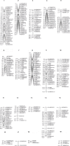
Fig 1
Fig 1. Genetic linkage map of Pseudocercospora fijiensis constructed from segregation data at 322 loci (233 DArT, 86 SSR and 3 minisatellite markers) among 135 individuals of a cross between the sequenced isolates CIRAD86 and CIRAD139A.
The Diversity Arrays Technology (DArT) markers were named according to the output of proprietary DArT analysis software. For each of the 19 linkage groups (listed on top) the cumulative map distances (cM) as calculated using the Haldane mapping function are shown to the left.
Fig 2
Fig 2. Phylogenetic analysis showing the placement of Dothideomycete species within the Capnodiales with expanded genomes.
At least two genome expansions may have taken place; one leading to the banana pathogen Pseudocercospora fijiensis and one that contributed to its close relative the tomato pathogen Cladosporium fulvum. Genome sizes and percentages of the genome containing repeat elements are indicated in parentheses.
Fig 3
Fig 3. Comparison of repeat classes among Zymoseptoria tritici, the only Dothideomycete with a completely sequenced genome, Pseudocercospora fijiensis and Cladosporium fulvum, the only other Dothideomycete known to have a transposon-expanded genome.
Fig 4
Fig 4. Repeat-induced point mutation (RIP) dinucleotide bias in Pseudocercospora fijiensis genome.
A clear CA <-> TA dinucleotide bias is observed in P. fijiensis repetitive families, indicating that RIP likely occurs and mutates CA nucleotide pairs to CT.
Fig 5
Fig 5. Comparison of the amount of repeat-induced point mutation (RIP) between AT-rich blocks and more GC-rich regions of the Pseudocercospora fijiensis genome as measured by the RIP index (CpA+TpG)/(ApC+GpT).
(A) AT-rich blocks have a lower RIP index indicating a depletion of RIP-susceptible sites due to a higher frequency of RIP compared to (B) an AT-poor region (higher GC) of the genome, which has a higher RIP index reflecting very little RIP. Four AT-rich blocks are shown along with one AT-poor region for comparison. Length of each block in kilobases is shown along the x-axis and the RIP index (CpA+TpG)/(ApC+GpT) is shown on the y-axis.
Fig 6
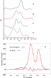
Fig 6. First-derivative graphs of melting curves of four different Dothideomycetes.
Examples of first-derivative graphs of melting curves obtained for Zymoseptoria tritici (A), Pseudocercospora fijiensis (B), P. eumusae (C) and P. musae (D). E: A plot of G+C contents from sequence reads of P. fijiensis. This graph is very similar to the melting-curve analyses showing the difference in G+C content between the genomes of P. fijiensis and the other banana pathogens versus the Z. tritici genome.
Fig 7
Fig 7. The numbers of long terminal repeat (LTR) retrotransposons in hypothetical age bins from less than one to more than 20 million years.
Estimated age of each transposon was calculated using the number of differences between its left and right repeats. These are considered identical at the time of insertion so all changes are likely due to mutations that occurred after transposition. All transition mutations were excluded to minimize the effects of repeat-induced point mutation.
Fig 8
Fig 8. Electrophoretic karyotypes of two strains of Pseudocercospora fijiensis.
A) Bands separated with conditions for small chromosomes. Lane 1, chromosomes from Saccharomyces cerevisiae as high-molecular-weight (HMW) marker; lane 2, strain CIRAD86; lane 3, strain E22. B) Bands separated under conditions to resolve medium and large chromosomes. Lane 1, chromosomes from Schizosaccharomyces pombe as HMW marker for large chromosomes; lane 2, strain CIRAD86; lane 3, strain E22; lane 4, chromosomes from Hansenula wingei as HMW marker for medium chromosomes in size. Marker sizes are in Kb.
Fig 9
Fig 9. Dot plot showing mesosynteny between the scaffolds of Pseudocercospora fijiensis and Dothistroma septosporum.
Fig 10
Fig 10. Genome-wide nucleotide comparison between Zymoseptoria tritici (lower half of the circle) and Pseudocercospora fijiensis (upper half of the circle).
The longest 28 scaffolds from P. fijiensis are shown. Gene content is conserved but is scattered among different chromosomes between these two fungi. There were no significant hits to dispensable chromosomes of Z. tritici (14–21). The 12 major scaffolds of P. fijiensis showing synteny are labeled in dark blue-green and the other 16 scaffolds are labeled in orange.
Fig 11
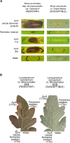
Fig 11. Infiltration of purified protein of the putative effector gene PfAvr4 from Pseudocercospora fijiensis into leaves of banana and tomato.
A: Infiltrations into leaves of resistant and susceptible banana varieties. B: Infiltrations into leaves of tomato with or without the Cf4 resistance gene known to interact with PfAVR4. Experiments were done with crude fermentor product and concentrated or diluted product. Fermentor medium alone and water were used as controls.

References

    1. Arzanlou M, Groenewald J, Fullerton R, Abeln E, Carlier J, Zapater MF, et al. Multiple gene genealogies and phenotypic characters differentiate several novel species of Mycosphaerella and related anamorphs on banana. Persoonia Mol Phylogeny Evol Fungi. 2008;20: 19. - PMC - PubMed
    1. Marin DH, Romero RA, Guzman M, Sutton TB. Black Sigatoka: An Increasing Threat to Banana Cultivation. Plant Dis. 2003;87: 208–222. - PubMed
    1. Crous PW, Mourichon X. Mycosphaerella eumusae and its anamorph Pseudocercospora eumusae spp. nov.: causal agent of eumusae leaf spot disease of banana. Sydowia. 2002;54: 35–43.
    1. Carlier J, Zapater MF, Lapeyre F, Jones DR, Mourichon X. Septoria leaf spot of banana: A newly discovered disease caused by Mycosphaerella eumusae (anamorph Septoria eumusae). Phytopathology. 2000;90: 884–890. 10.1094/PHYTO.2000.90.8.884 - DOI - PubMed
    1. Carlier J, Lebrun MH, Zapater MF, Dubois C, Mourichon X. Genetic structure of the global population of banana black leaf streak fungus, Mycosphaerella fijiensis. Mol Ecol. 1996;5: 499–510. 10.1111/j.1365-294X.1996.tb00342.x - DOI

Publication types