. 2016 Dec;44(12):e1236-e1245.
doi: 10.1097/CCM.0000000000001999.

Carbon Monoxide Improves Efficacy of Mesenchymal Stromal Cells During Sepsis by Production of Specialized Proresolving Lipid Mediators

Affiliations

Carbon Monoxide Improves Efficacy of Mesenchymal Stromal Cells During Sepsis by Production of Specialized Proresolving Lipid Mediators

Konstantin Tsoyi et al. Crit Care Med. 2016 Dec.

Abstract

Objectives: Mesenchymal stromal cells are being investigated as a cell-based therapy for a number of disease processes, with promising results in animal models of systemic inflammation and sepsis. Studies are ongoing to determine ways to further improve the therapeutic potential of mesenchymal stromal cells. A gas molecule that improves outcome in experimental sepsis is carbon monoxide. We hypothesized that preconditioning of mesenchymal stromal cells with carbon monoxide ex vivo would promote further therapeutic benefit when cells are administered in vivo after the onset of polymicrobial sepsis in mice.

Design: Animal study and primary cell culture.

Setting: Laboratory investigation.

Subjects: BALB/c mice.

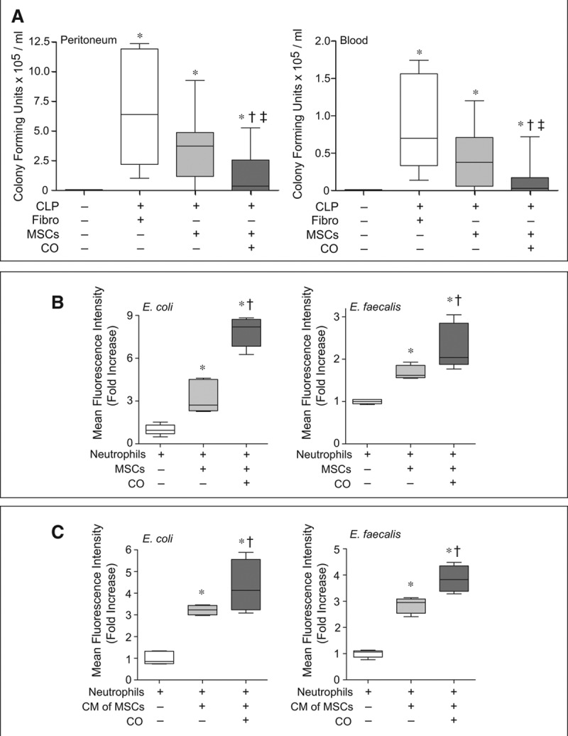
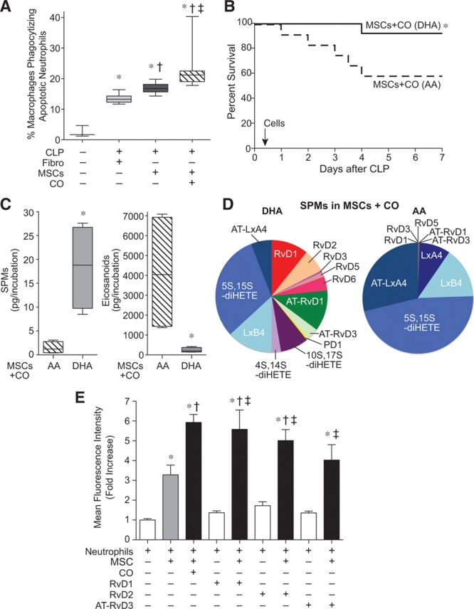
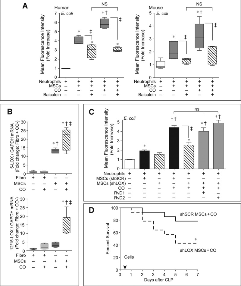
Interventions: Polymicrobial sepsis was induced by cecal ligation and puncture. Mesenchymal stromal cells, mesenchymal stromal cells-conditioned with carbon monoxide, fibroblasts, or fibroblasts-conditioned with carbon monoxide were delivered by tail vein injections to septic mice. The mice were assessed for survival, bacterial clearance, and the inflammatory response during sepsis in each of the groups. Mesenchymal stromal cells were also assessed for their ability to promote bacterial phagocytosis by neutrophils, the production of specialized proresolving lipid mediators, and their importance for mesenchymal stromal cells function using gene silencing.

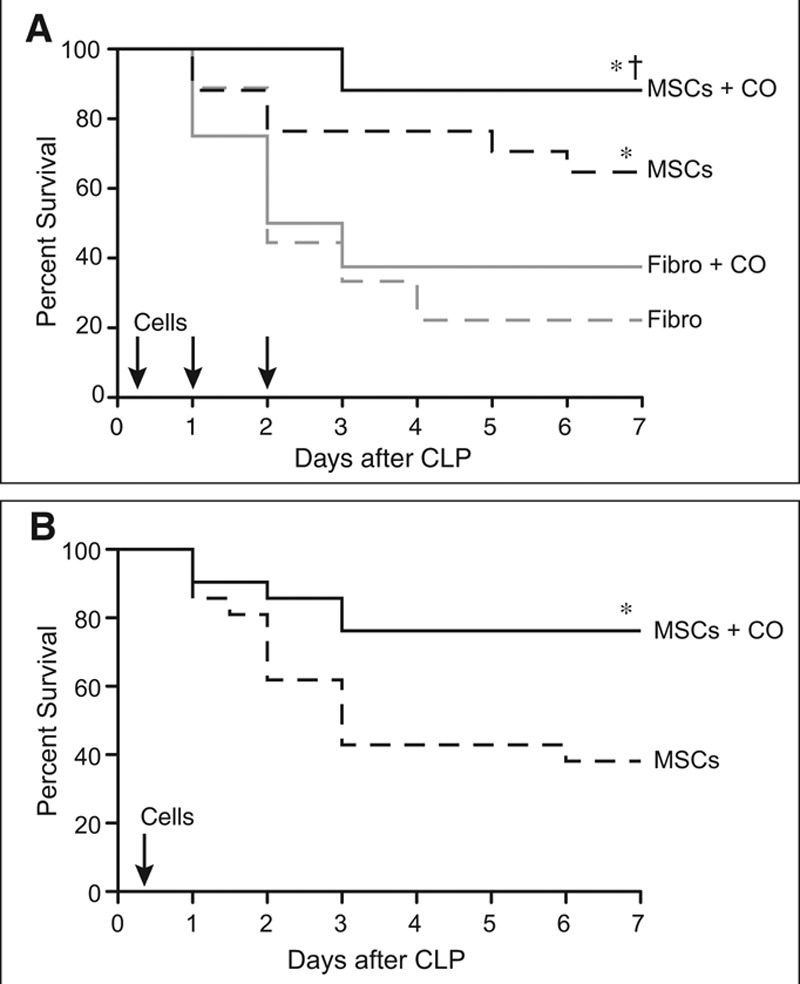
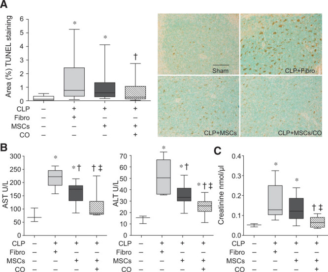
Measurements and main results: Ex vivo preconditioning with carbon monoxide allowed mesenchymal stromal cells to be administered later after the onset of sepsis (6 hr), and yet maintain their therapeutic effect with increased survival. Carbon monoxide preconditioned mesenchymal stromal cells were also able to alleviate organ injury, improve bacterial clearance, and promote the resolution of inflammation. Mesenchymal stromal cells exposed to carbon monoxide, with docosahexaenoic acid substrate, produced specialized proresolving lipid mediators, particularly D-series resolvins, which promoted survival. Silencing of lipoxygenase pathways (5-lipoxygenase and 12/15-lipoxygenase), which are important enzymes for specialized proresolving lipid mediator biosynthesis, resulted in a loss of therapeutic benefit bestowed on mesenchymal stromal cells by carbon monoxide.

Conclusions: Taken together, these data suggest that production of specialized proresolving lipid mediators contribute to improved mesenchymal stromal cell efficacy when exposed to carbon monoxide, resulting in an improved therapeutic response during sepsis.


Conflict of interest statement

Dr. Perrella received support for article research from the National Institutes of Health (NIH). His institution received funding from the NIH (National Heart, Lung, and Blood Institute [NHLBI]). Dr. Tsoyi received support for article research from the NIH. His institution received funding from the NIH. Drs. Dalli and Colas received support for article research from the NIH. Drs. Ghanta and Ith received support for article research from the NIH. Their institutions received funding from the NIH. Dr. Fredenburgh received support for article research from the NIH. Her institution received funding from the NHLBI. Drs. Baron and Choi received support for article research from the NIH. Dr. Serhan received support for article research from the NIH and disclosed other support (SAB member Corbus, SAB member Inflammation Foundation, SAB Solutex. He is an inventor on patents [resolvins] assigned to BWH and licensed to Resolvyx Pharmaceuticals. He is a scientific founder of Resolvyx Pharmaceuticals and owns equity in the company [no current relationship but still hold founder stock]. His interests were reviewed and are managed by the Brigham and Women’s Hospital and Partners HealthCare in accordance with their conflict of interest policies). Dr. Liu received support for article research from the NIH and the AHA. Her institution received funding from the NIH and the AHA. The remaining authors have disclosed that they do not have any potential conflicts of interest.

Figures

Figure 1.
Figure 1.
Administration of mesenchymal stromal cells (MSCs) preconditioned with carbon monoxide (CO) improves efficacy of the cells and survival in cecal ligation and puncture (CLP)-induced sepsis. A, BALB/c mice were randomly separated into four groups: fibroblasts (Fibro), dashed gray line, n = 9; Fibro+CO, solid gray line, n = 8; MSCs, dashed black line, n = 17; and MSCs+CO, solid black line, n = 17. All mice were subjected to CLP, and 2 hr later the mice were treated with 5 × 105 cells by tail vein injection. Treatments were repeated at 24 and 48 hr after surgery with 2.5 × 105 cells. Survival of mice was monitored for 7 d, and data are presented as a Kaplan-Meier survival curve, and analyzed by log-rank test. * MSCs versus Fibro, p = 0.034; * MSCs+CO versus Fibro, p = 0.0002; and † MSCs+CO versus Fibro+CO, p = 0.003. B, BALB/c mice were randomly separated into two groups: MSCs, dashed black line, n = 21; and MSCs+CO, solid black line, n = 21. All mice were subjected to CLP, and after 6 hr the mice were treated with 5 × 105 cells by tail vein injection. Survival of mice and data analysis as described in (A). *MSCs+CO versus MSCs, p = 0.016.
Figure 2.
Figure 2.
Administration of mesenchymal stromal cells (MSCs) preconditioned with carbon monoxide (CO) decreases tissue injury in cecal ligation and puncture (CLP)-induced sepsis. Mice were subjected to sham or CLP surgery (CLP+fibroblasts [Fibro], CLP+MSCs, CLP+MSCs/CO). A total of 5 × 105 cells were administered by tail vein injection 6 hr after CLP. At 24 hr, mice were anesthetized, and blood and spleens were harvested. A, Spleen injury was evaluated by terminal deoxynucleotidyl transferase dUTP nick end labeling (TUNEL) staining (brown staining, right), and quantitated (left). The bar in the sham represents a length of 50 μm. Data are presented as box plots, which show median values and interquartile ranges. Analysis was done by Kruskal-Wallis testing (also used in B and C). Sham, white bar, n = 3; CLP+Fibro, light gray bar, n = 7; CLP+MSCs, dark gray bar, n = 8; CLP+MSCs/CO, striped bar, n = 8. p = 0.0001, with significant comparisons * versus sham and † versus CLP+Fibro. B, Plasma aspartate aminotransferase (AST) and alanine aminotransferase (ALT) data are shown as box plots. Sham, n = 3; CLP+Fibro, light gray bar, n = 6; CLP+MSCs, dark gray bar, n = 8; CLP+MSCs/CO, striped bar, n = 9. p = 0.002 and p = 0.0008, respectively, with significant comparisons * versus sham, † versus CLP+Fibro, and ‡ versus CLP+MSCs. C, Plasma creatinine levels are shown as box plots. Sham, n = 3; CLP+Fibro, light gray bar, n = 6; CLP+MSCs, dark gray bar, n = 6; CLP+MSCs/CO, striped bar, n = 5. p = 0.018, and significant comparisons of specific groups * versus sham, † versus CLP+Fibro, and ‡ versus CLP+MSCs.
Figure 3.
Figure 3.
Administration of mesenchymal stromal cells (MSCs) preconditioned with carbon monoxide (CO) improves bacterial clearance in cecal ligation and puncture (CLP)-induced sepsis and increases bacterial phagocytosis by neutrophils. A, Mice were subjected to sham or CLP surgery (CLP+fibroblasts [Fibro], CLP+MSCs, CLP+MSCs/CO). A total of 5 × 105 cells were administered by tail vein injection 6 hr after CLP. At 24 hr, peritoneal fluid (left) and blood samples (right) were collected and analyzed. Data are presented as box plots, which show median values and interquartile ranges. Analysis was done by Kruskal-Wallis testing (also used in B and C). Left, sham, n = 11; CLP+Fibro, white box, n = 6; CLP+MSCs, light gray box, n = 9; CLP+MSCs/CO, dark gray box, n = 12. Right, sham, n = 7; CLP+Fibro, white box, n = 6; CLP+MSCs, light gray box, n = 10; CLP+MSCs/CO, dark gray box, n = 12. p < 0.0001 left and p = 0.001 right, with significant comparisons * versus sham, † versus CLP+Fibro, and ‡ versus CLP+MSCs. B, Isolated neutrophils were incubated with green fluorescent protein (GFP)-labeled E. coli or E. faecalis in the presence of MSCs (light gray bars), MSCs+CO (dark gray bars) or in the absence of MSCs (white bars). Data are presented as box plots, n = 5–7 per groups from three independent experiments. p < 0.0008 for E. coli and p = 0.0022 for E. faecalis, with significant comparisons * versus no MSCs, and † versus MSCs. C, Isolated neutrophils were incubated with GFP-labeled E. coli or E. faecalis with addition of conditioned medium (CM) from MSCs (light gray bars), MSCs+CO (dark gray bars), or basal medium (white bars). Data are presented as box plots, n = 4–5 per group from three independent experiments. p < 0.0064 for E. coli and p < 0.0047 for E. faecalis, with significant comparisons * versus basal medium, and † versus CM of MSCs.
Figure 4.
Figure 4.
Carbon monoxide (CO) preconditioning induces production of specialized proresolving lipid mediators (SPMs) in mesenchymal stromal cells (MSCs), which mimic the CO effect by enhancing neutrophil phagocytosis of bacteria. A, Mice were subjected to sham or cecal ligation and puncture (CLP) surgery (CLP+fibroblasts [Fibro], CLP+MSCs, CLP+MSCs/CO). A total of 5 × 105 cells were administered by tail vein injection 6 hr after CLP. At 24 hr, peritoneal fluid was collected and efferocytosis assays were performed. Data are presented as box plots, which show median values and interquartile ranges. Sham, n = 3; CLP+Fibro, light gray bar, n = 6; CLP+MSCs, dark gray bar, n = 6; CLP+MSCs/CO, striped bar, n = 7. Analysis was done by Kruskal-Wallis testing (p = 0.0004), with significant comparisons * versus sham, † versus CLP+Fibro, and ‡ versus CLP+MSCs. B, Mice were subjected to CLP, and 6 hr later, the mice received 5 × 105 MSCs treated with CO in the presence of docosahexaenoic acid (DHA) (10 μM), solid black line, n = 12 or MSCs-treated with CO in the presence of arachidonic acid (AA) (10 μM), dashed black line, n = 12. Survival of mice was monitored for 7 d and data are presented as a Kaplan-Meier survival curve, and analyzed by log-rank test. *versus MSCs+CO (AA), p = 0.040. C, Human MSCs were exposed to CO in the presence of DHA or AA, followed by lipid mediator profiling. Total SPMs are shown left, and total eicosanoids right. Data are presented as box plots, n = 4 samples per group from two independent experiments. Analysis was done by Mann-Whitney U testing. * versus MSCs+CO in the presence of AA (p = 0.03 for SPMs and p = 0.03 for eicosanoids). D, Representative pie charts of SPM levels in MSCs conditioned with CO, in the presence of DHA or AA. E, Human neutrophils were incubated with green fluorescent protein-labeled E. coli alone (white bars, n = 16), in the presence of MSCs (gray bar, n = 16), or in the presence of CO-conditioned MSCs (black bar, n = 16) or MSCs conditioned with resolvin D1 (RvD1, 10 nM), Rv D2 (10 nM), or aspirin-triggered Rv D3 (AT-RvD3, 10 nM) (black bars, n = 8). Data are presented as fold increase in mean fluorescence intensity, mean ± sem, from three independent experiments. Analysis was done by one-way analysis of variance (p < 0.0001), with significant comparisons * versus no MSCs, † versus MSCs, and ‡ versus its RvD control. HETE = hydroxyeicosatetraenoic acid, Lx = lipoxin, PD1 = Protectin D1.
Figure 5.
Figure 5.
Lipoxygenase pathways contribute to the carbon monoxide (CO)-mediated effects of mesenchymal stromal cells (MSCs) on neutrophil phagocytosis, and silencing of 5-lipoxygenase (LOX) and 12/15-LOX results in a loss of improved MSC efficacy by CO conditioning. A, MSCs were pretreated with baicalein (10 μM) or vehicle, and then cocultured with human (left) or mouse (right) neutrophils in the presence or absence of green fluorescent protein (GFP)-labeled E. coli. Data are presented as box plots, which show median values and interquartile ranges. Analysis was done by Kruskal-Wallis testing (also used in B). n = 4–5 per group from three independent experiments. p = 0.0016 and p = 0.0062, human and mouse, respectively, and significant comparisons of specific groups * versus no MSCs, † versus MSCs, and ‡ between designated groups. B, Fibroblasts (Fibro) and MSCs were incubated with CO or ambient air for 4 hr, and then RNA was extracted and quantitative real-time polymerase chain reaction performed for 5-LOX (upper) and 12/15-LOX (lower). Data are presented as box plots, n = 4–6 per group from three independent experiments. p = 0.0008 for 5-LOX and 12/15-LOX, with significant comparisons * versus Fibro, † versus Fibro+CO, and ‡ versus MSCs. C, MSCs were silenced with scrambled (SCR), or both 5-LOX and 12/15-LOX = small hairpin RNAs (shRNAs) (method used to silence 5-LOX and 12/15 LOX (shLOX [defined in the narrative of the article]). The MSCs were exposed to CO or ambient air, and then cocultured with neutrophils in the presence of GFP-labeled E. coli. shLOX MSCs exposed to CO were also exposed to resolvin D1 (RvD1, 10 nM) or Rv D2 (10 nM). Data are presented as fold increase in mean fluorescence intensity, mean ± sem, n = 6–12 per group from three independent experiments. Analysis was done by one-way analysis of variance (p < 0.0001) with significant comparisons * versus no MSCs, † versus MSCs (SCR shRNA sequence [shSCR]), and ‡ between designated groups. D, BALB/c mice were randomly separated into two groups, shSCR MSCs+CO (solid black line, n = 14) or shLOX MSCs+CO (dashed black line, n = 14). Six hours after cecal ligation and puncture (CLP), the mice were treated with 5 × 105 cells by tail vein injection. Animal survival was monitored for 7 d, and data are presented as a Kaplan-Meier survival curve, and analyzed by log-rank test. * versus shSCR MSCs+CO, p = 0.049. GAPDH = glyceraldehyde-3-phosphate dehydrogenase, mRNA = messenger RNA, NS = not significant.

Comment in

References

    1. Annane D, Bellissant E, Cavaillon JM. Septic shock. Lancet. 2005;365:63–78. - PubMed
    1. Hotchkiss RS, Karl IE. The pathophysiology and treatment of sepsis. N Engl J Med. 2003;348:138–150. - PubMed
    1. Marik PE. Surviving sepsis: Going beyond the guidelines. Ann Intensive Care. 2011;1:17. - PMC - PubMed
    1. Opal SM, Dellinger RP, Vincent JL, et al. The next generation of sepsis clinical trial designs: What is next after the demise of recombinant human activated protein C?. Crit Care Med. 2014;42:1714–1721. - PMC - PubMed
    1. Serhan CN, Chiang N, Van Dyke TE. Resolving inflammation: Dual anti-inflammatory and pro-resolution lipid mediators. Nat Rev Immunol. 2008;8:349–361. - PMC - PubMed