

MicroRNA-21 Increases Proliferation and Cisplatin Sensitivity of Osteosarcoma-Derived Cells
- PMID: 27513462
- PMCID: PMC4981312
- DOI: 10.1371/journal.pone.0161023
MicroRNA-21 Increases Proliferation and Cisplatin Sensitivity of Osteosarcoma-Derived Cells
Abstract
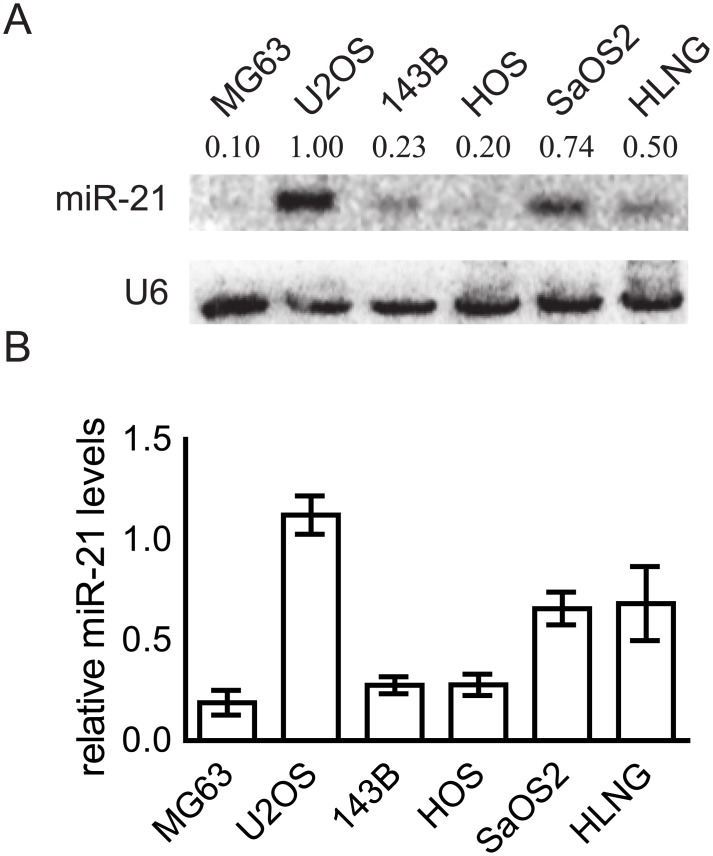
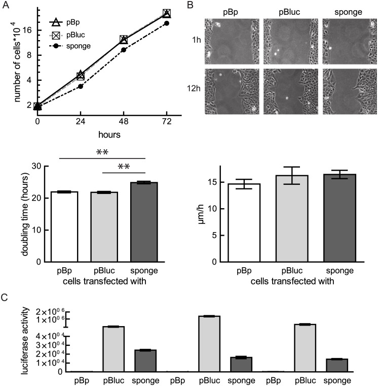
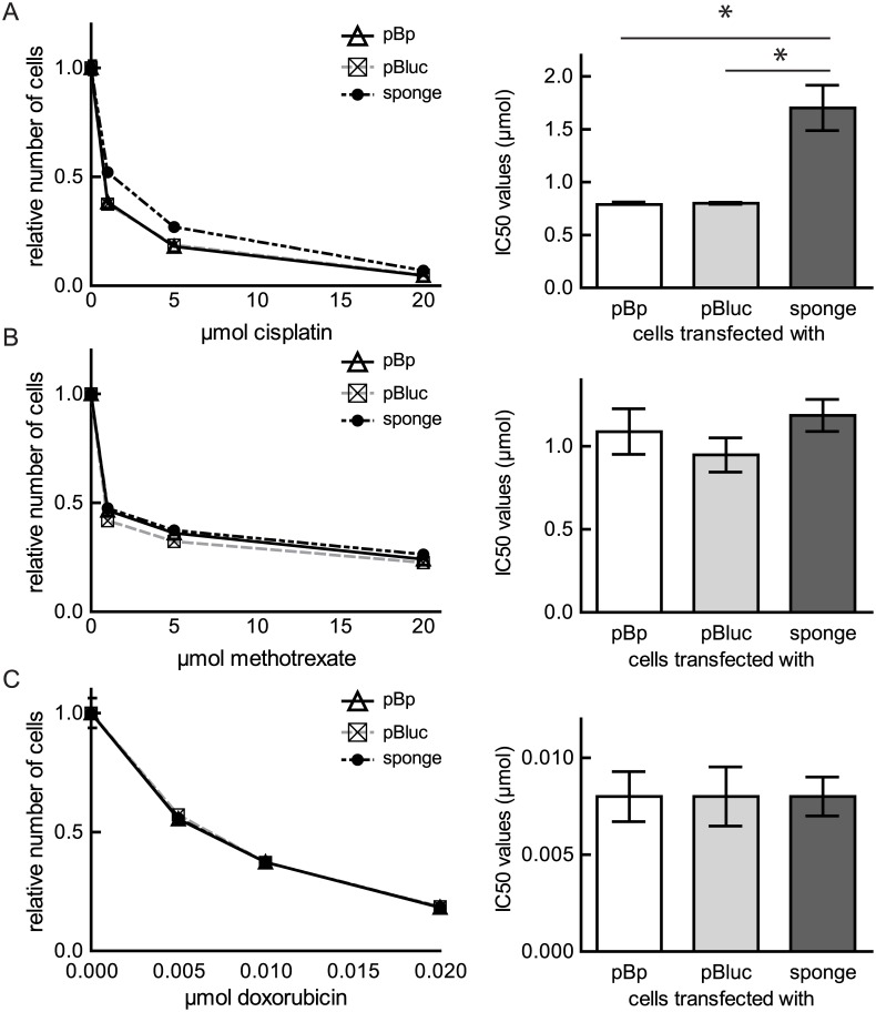
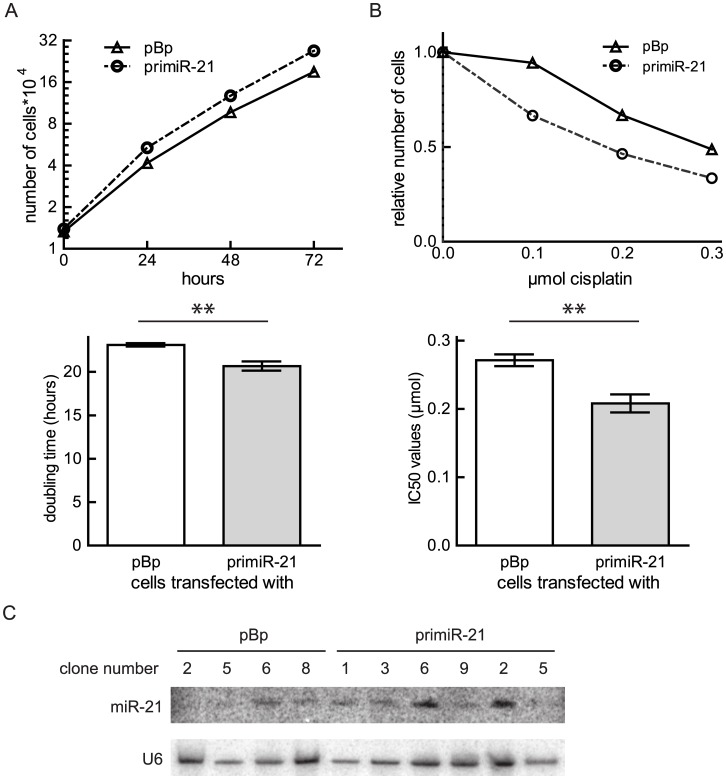
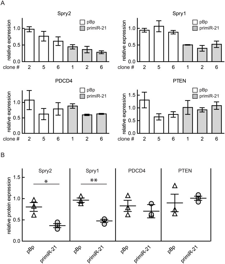
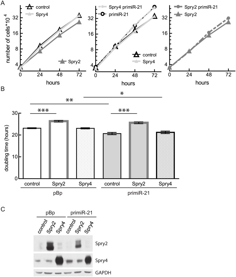
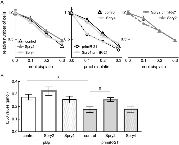
Osteosarcoma is the most common primary bone tumor and poor prognosis for osteosarcoma patients is mainly due to chemotherapy resistance. MicroRNAs are important to maintain pathophysiological mechanisms of cancer and influence cell sensitivity to chemotherapy. In this study, we tested the functions of microRNA-21 for malignant features as well as for drug resistance of osteosarcoma. We used Northern blot to measure microRNA-21 levels in osteosarcoma-derived cell lines. MicroRNA-21 activity was modulated by either expressing a sponge to decrease its activity in an osteosarcoma-derived cell line expressing high levels of microRNA-21 or by introducing pri-microRNA-21 in a cell line with low endogenous levels. Cell migration was determined in a scratch assay and cell proliferation was measured by performing growth curve analysis. Sensitivity of the cells towards chemotherapeutics was investigated by performing cell viability assays and calculating the IC50 values. While cell migration was unaffected by modulated microRNA-21 levels, microRNA-21 inhibition slowed proliferation and exogenously expressed microRNA-21 promoted this process. Modulated microRNA-21 activity failed to effect sensitivity of osteosarcoma-derived cell lines to doxorubicin or methotrexate. Contrarily, reduction of microRNA-21 activity resulted in enhanced resistance towards cisplatin while ectopic expression of microRNA-21 showed the opposite effect. Increased microRNA-21 levels repressed the expression of Sprouty2 and ectopic expression of Sprouty2 was able to largely rescue the observed effects of microRNA-21 in osteosarcoma. In summary, our data indicate that in osteosarcoma microRNA-21 expression is an important component for regulation of cell proliferation and for determining sensitivity to cisplatin.
Conflict of interest statement
Figures
Similar articles
-
Circular RNA LARP4 correlates with decreased Enneking stage, better histological response, and prolonged survival profiles, and it elevates chemosensitivity to cisplatin and doxorubicin via sponging microRNA-424 in osteosarcoma.J Clin Lab Anal. 2020 Feb;34(2):e23045. doi: 10.1002/jcla.23045. Epub 2019 Oct 22. J Clin Lab Anal. 2020. PMID: 31642110 Free PMC article.
-
MicroRNA‑22 inhibits the proliferation and migration, and increases the cisplatin sensitivity, of osteosarcoma cells.Mol Med Rep. 2018 May;17(5):7209-7217. doi: 10.3892/mmr.2018.8790. Epub 2018 Mar 20. Mol Med Rep. 2018. PMID: 29568877 Free PMC article.
-
Hypoxia-inducible microRNA-488 regulates apoptosis by targeting Bim in osteosarcoma.Cell Oncol (Dordr). 2016 Oct;39(5):463-471. doi: 10.1007/s13402-016-0288-2. Epub 2016 Jul 4. Cell Oncol (Dordr). 2016. PMID: 27376839
-
microRNA and Bone Cancer.Adv Exp Med Biol. 2015;889:201-30. doi: 10.1007/978-3-319-23730-5_11. Adv Exp Med Biol. 2015. PMID: 26659003 Review.
-
MicroRNAs as the pivotal regulators of cisplatin resistance in osteosarcoma.Pathol Res Pract. 2023 Sep;249:154743. doi: 10.1016/j.prp.2023.154743. Epub 2023 Aug 6. Pathol Res Pract. 2023. PMID: 37549518 Review.
Cited by
-
Circular RNA LARP4 correlates with decreased Enneking stage, better histological response, and prolonged survival profiles, and it elevates chemosensitivity to cisplatin and doxorubicin via sponging microRNA-424 in osteosarcoma.J Clin Lab Anal. 2020 Feb;34(2):e23045. doi: 10.1002/jcla.23045. Epub 2019 Oct 22. J Clin Lab Anal. 2020. PMID: 31642110 Free PMC article.
-
Biomarker significance of plasma and tumor miR-21, miR-221, and miR-106a in osteosarcoma.Oncotarget. 2017 May 27;8(57):96738-96752. doi: 10.18632/oncotarget.18236. eCollection 2017 Nov 14. Oncotarget. 2017. PMID: 29228567 Free PMC article.
-
An overview of resistance to chemotherapy in osteosarcoma and future perspectives.Cancer Drug Resist. 2022 Jun 23;5(3):762-793. doi: 10.20517/cdr.2022.18. eCollection 2022. Cancer Drug Resist. 2022. PMID: 36176756 Free PMC article. Review.
-
Non-coding RNAs in cancer: platforms and strategies for investigating the genomic "dark matter".J Exp Clin Cancer Res. 2020 Jun 20;39(1):117. doi: 10.1186/s13046-020-01622-x. J Exp Clin Cancer Res. 2020. PMID: 32563270 Free PMC article. Review.
-
Long noncoding RNA DNAJC3-AS1 promotes osteosarcoma progression via its sense-cognate gene DNAJC3.Cancer Med. 2019 Feb;8(2):761-772. doi: 10.1002/cam4.1955. Epub 2019 Jan 16. Cancer Med. 2019. PMID: 30652414 Free PMC article.
References
MeSH terms
Substances
LinkOut - more resources
Full Text Sources
Other Literature Sources
Medical
