

Reproducible Construction of Surface Tension-Mediated Honeycomb Concave Microwell Arrays for Engineering of 3D Microtissues with Minimal Cell Loss
- PMID: 27513567
- PMCID: PMC4981302
- DOI: 10.1371/journal.pone.0161026
Reproducible Construction of Surface Tension-Mediated Honeycomb Concave Microwell Arrays for Engineering of 3D Microtissues with Minimal Cell Loss
Abstract
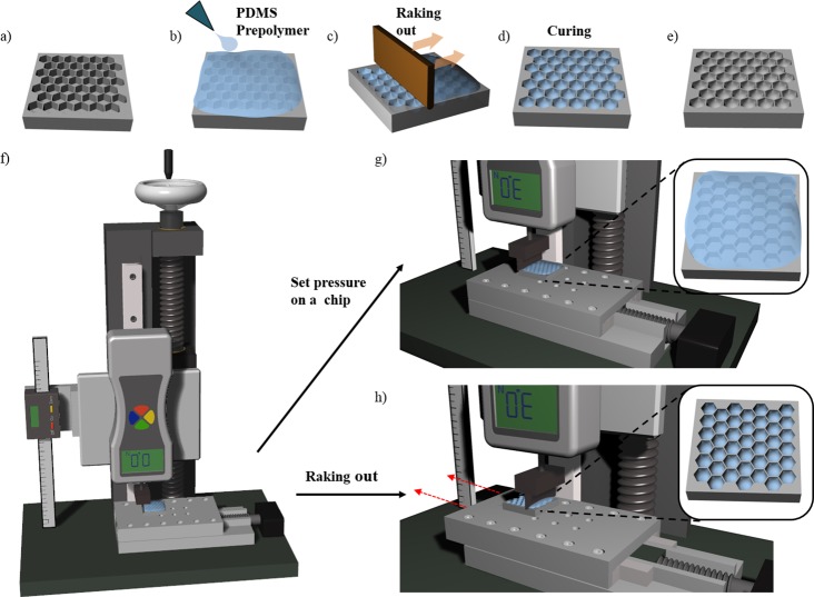
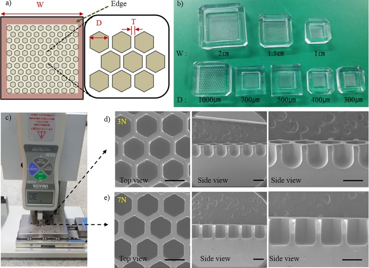
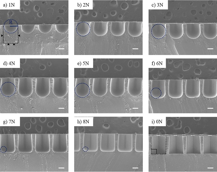
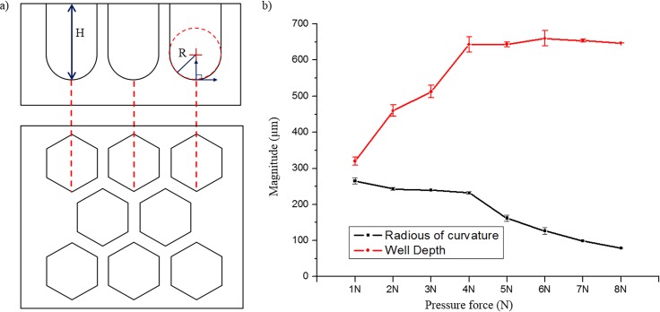
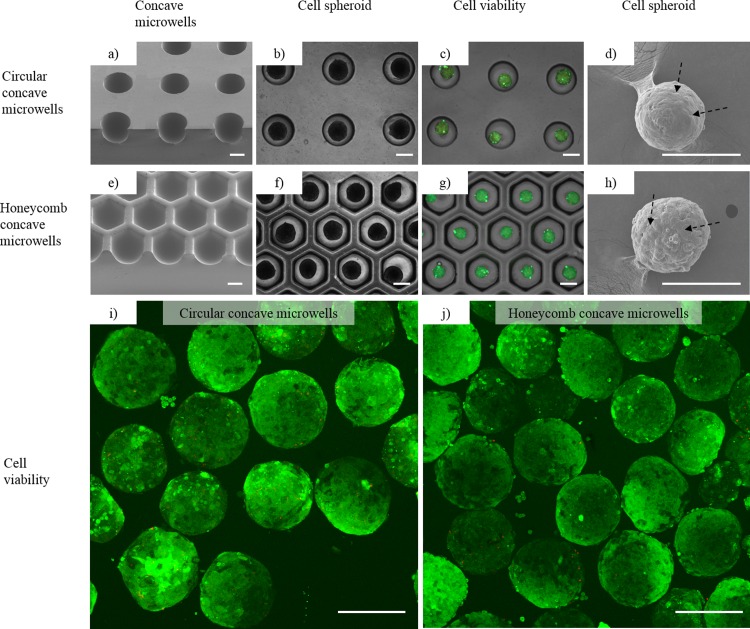
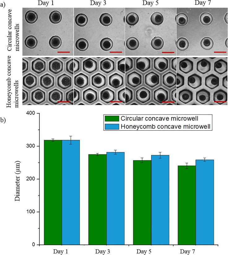
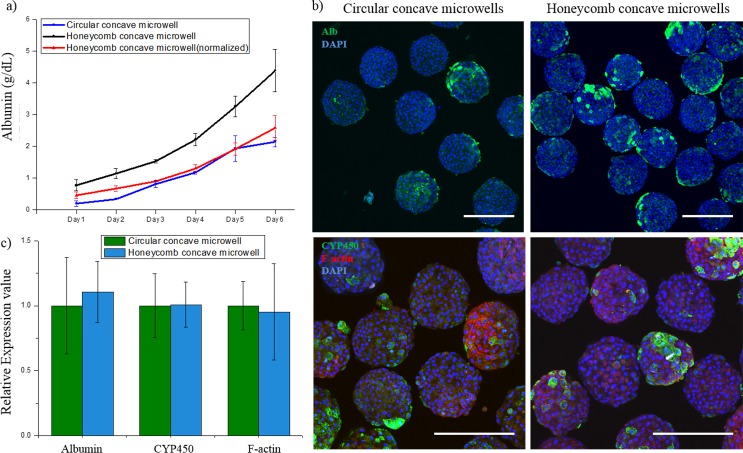
The creation of engineered 3D microtissues has attracted prodigious interest because of the fact that this microtissue structure is able to mimic in vivo environments. Such microtissues can be applied extensively in the fields of regenerative medicine and tissue engineering, as well as in drug and toxicity screening. Here, we develop a novel method of fabricating a large number of dense honeycomb concave microwells via surface tension-mediated self-construction. More specifically, in order to control the curvature and shape of the concavity in a precise and reproducible manner, a custom-made jig system was designed and fabricated. By applying a pre-set force using the jig system, the shape of the honeycomb concave well was precisely and uniformly controlled, despite the fact that wells were densely packed. The thin wall between the honeycomb wells enables the minimization of cell loss during the cell-seeding process. To evaluate the performance of the honeycomb microwell array, rat hepatocytes were seeded, and spheroids were successfully formed with uniform shape and size. Liver-specific functions such as albumin secretion and cytochrome P450 were subsequently analyzed. The proposed method of fabricating honeycomb concave wells is cost-effective, simple, and reproducible. The honeycomb well array can produce multiple spheroids with minimal cell loss, and can lead to significant contributions in tissue engineering and organ regeneration.
Conflict of interest statement
Figures
Similar articles
-
Networked concave microwell arrays for constructing 3D cell spheroids.Biofabrication. 2017 Nov 30;10(1):015001. doi: 10.1088/1758-5090/aa9876. Biofabrication. 2017. PMID: 29190216
-
Enhanced oxygen permeability in membrane-bottomed concave microwells for the formation of pancreatic islet spheroids.Acta Biomater. 2018 Jan;65:185-196. doi: 10.1016/j.actbio.2017.10.045. Epub 2017 Oct 31. Acta Biomater. 2018. PMID: 29101017
-
Fabrication of agarose concave petridish for 3D-culture microarray method for spheroids formation of hepatic cells.J Mater Sci Mater Med. 2018 Apr 19;29(5):49. doi: 10.1007/s10856-018-6058-0. J Mater Sci Mater Med. 2018. PMID: 29675647
-
Scaffold-free microtissues: differences from monolayer cultures and their potential in bone tissue engineering.Clin Oral Investig. 2013 Jan;17(1):9-17. doi: 10.1007/s00784-012-0763-8. Epub 2012 Jun 14. Clin Oral Investig. 2013. PMID: 22695872 Free PMC article. Review.
-
Bottom-Up Engineering of Well-Defined 3D Microtissues Using Microplatforms and Biomedical Applications.Adv Healthc Mater. 2016 Jan 7;5(1):56-74. doi: 10.1002/adhm.201500107. Epub 2015 Apr 16. Adv Healthc Mater. 2016. PMID: 25880830 Review.
Cited by
-
Bioprinting of a Hepatic Tissue Model Using Human-Induced Pluripotent Stem Cell-derived Hepatocytes for Drug-Induced Hepatotoxicity Evaluation.Int J Bioprint. 2022 Jun 14;8(3):581. doi: 10.18063/ijb.v8i3.581. eCollection 2022. Int J Bioprint. 2022. PMID: 36105133 Free PMC article.
-
Challenges of applying multicellular tumor spheroids in preclinical phase.Cancer Cell Int. 2021 Mar 4;21(1):152. doi: 10.1186/s12935-021-01853-8. Cancer Cell Int. 2021. PMID: 33663530 Free PMC article. Review.
-
Fabrication approaches for high-throughput and biomimetic disease modeling.Acta Biomater. 2021 Sep 15;132:52-82. doi: 10.1016/j.actbio.2021.03.006. Epub 2021 Mar 11. Acta Biomater. 2021. PMID: 33716174 Free PMC article. Review.
-
A Highly Reproducible Micro U-Well Array Plate Facilitating High-Throughput Tumor Spheroid Culture and Drug Assessment.Glob Chall. 2020 Nov 4;5(2):2000056. doi: 10.1002/gch2.202000056. eCollection 2021 Feb. Glob Chall. 2020. PMID: 33552551 Free PMC article.
-
Fabrication of Concave Microwells and Their Applications in Micro-Tissue Engineering: A Review.Micromachines (Basel). 2022 Sep 19;13(9):1555. doi: 10.3390/mi13091555. Micromachines (Basel). 2022. PMID: 36144178 Free PMC article. Review.
References
MeSH terms
LinkOut - more resources
Full Text Sources
Other Literature Sources
