

D-Dimer Levels Predict Myocardial Injury in ST-Segment Elevation Myocardial Infarction: A Cardiac Magnetic Resonance Imaging Study
- PMID: 27513758
- PMCID: PMC4981325
- DOI: 10.1371/journal.pone.0160955
D-Dimer Levels Predict Myocardial Injury in ST-Segment Elevation Myocardial Infarction: A Cardiac Magnetic Resonance Imaging Study
Abstract
Objectives: Elevated D-dimer levels on admission predict prognosis in patients undergoing primary percutaneous coronary intervention (PCI) for ST-segment elevation myocardial infarction (STEMI), but the association of D-dimer levels with structural markers of myocardial injury in these patients is unknown.
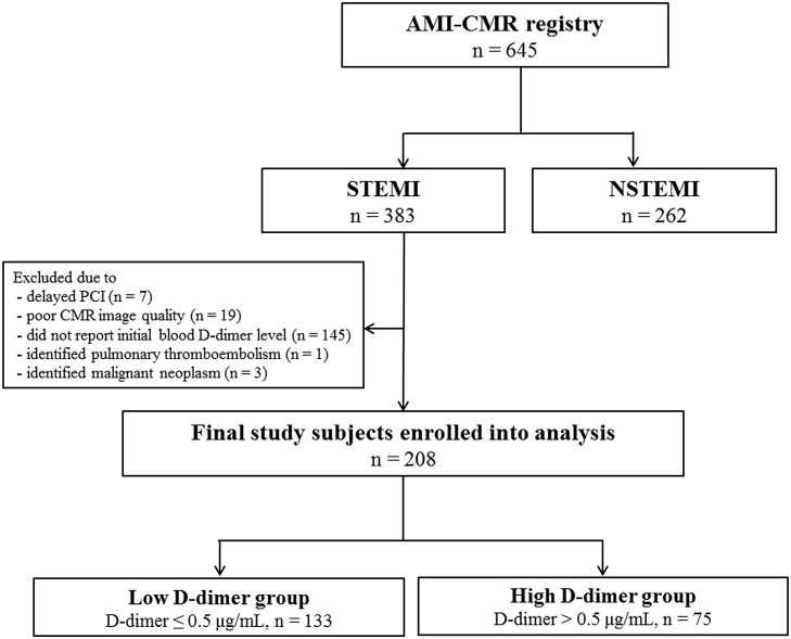
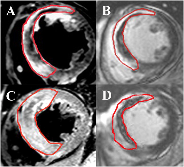
Methods: We performed cardiac magnetic resonance (CMR) imaging in 208 patients treated with primary PCI for STEMI. CMR was performed a median of 3 days after the index procedure. Of the 208 patients studied, 75 patients had D-dimer levels above the normal range on admission (>0.5 μg/mL; high D-dimer group) while 133 had normal levels (≤0.5 μg/mL; low D-dimer group). The primary outcome was myocardial infarct size assessed by CMR. Secondary outcomes included area at risk (AAR), microvascular obstruction (MVO) area, and myocardial salvage index (MSI).
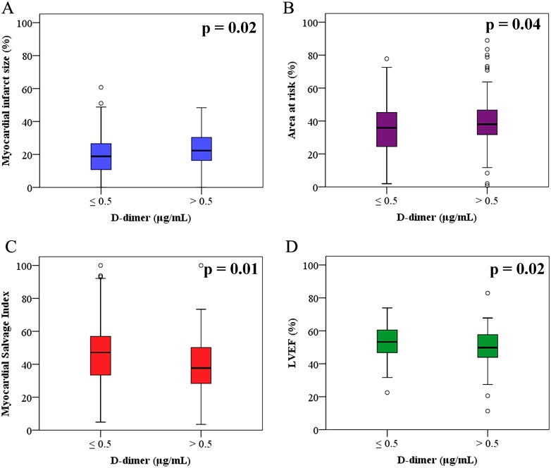
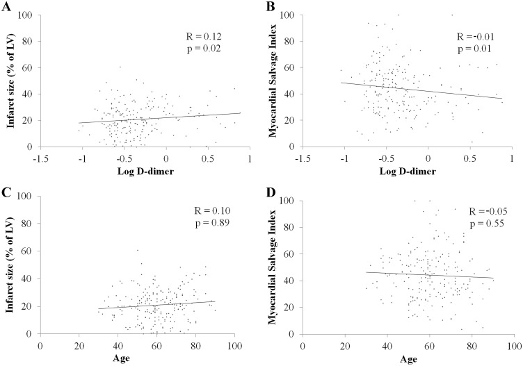
Results: In CMR analysis, myocardial infarct size was larger in the high D-dimer group than in the low D-dimer group (22.3% [16.2-30.5] versus 18.8% [10.7-26.7]; p = 0.02). Compared to the low D-dimer group, the high D-dimer group also had a larger AAR (38.1% [31.7-46.9] versus 35.8% [24.2-45.3]; p = 0.04) and a smaller MSI (37.7 [28.2-46.9] versus 47.1 [33.2-57.0]; p = 0.01). In multivariate analysis, high D-dimer levels were significantly associated with larger myocardial infarct (OR 2.59; 95% CI 1.37-4.87; p<0.01) and lower MSI (OR 2.62; 95% CI 1.44-4.78; p<0.01).
Conclusions: In STEMI patients undergoing primary PCI, high D-dimer levels on admission were associated with a larger myocardial infarct size, a greater extent of AAR, and lower MSI, as assessed by CMR data. Elevated initial D-dimer level may be a marker of advanced myocardial injury in patients treated with primary PCI for STEMI.
Conflict of interest statement
Figures
References
-
- Dunn KL, Wolf JP, Dorfman DM, Fitzpatrick P, Baker JL, Goldhaber SZ. Normal D-dimer levels in emergency department patients suspected of acute pulmonary embolism. Journal of the American College of Cardiology. 2002;40(8):1475–8. Epub 2002/10/24. . - PubMed
-
- Oldgren J, Linder R, Grip L, Siegbahn A, Wallentin L. Coagulation activity and clinical outcome in unstable coronary artery disease. Arteriosclerosis, thrombosis, and vascular biology. 2001;21(6):1059–64. Epub 2001/06/09. . - PubMed
-
- Ridker PM, Hennekens CH, Cerskus A, Stampfer MJ. Plasma concentration of cross-linked fibrin degradation product (D-dimer) and the risk of future myocardial infarction among apparently healthy men. Circulation. 1994;90(5):2236–40. Epub 1994/11/01. . - PubMed
-
- Menown IB, Mathew TP, Gracey HM, Nesbitt GS, Murray P, Young IS, et al. Prediction of Recurrent Events by D-Dimer and Inflammatory Markers in Patients with Normal Cardiac Troponin I (PREDICT) Study. American heart journal. 2003;145(6):986–92. Epub 2003/06/11. 10.1016/s0002-8703(03)00169-8 . - DOI - PubMed
MeSH terms
Substances
LinkOut - more resources
Full Text Sources
Other Literature Sources
Medical
Miscellaneous
