. 2016 Aug 11;11(8):e0160622.
doi: 10.1371/journal.pone.0160622. eCollection 2016.

Silica-Triggered Autoimmunity in Lupus-Prone Mice Blocked by Docosahexaenoic Acid Consumption

Affiliations

Silica-Triggered Autoimmunity in Lupus-Prone Mice Blocked by Docosahexaenoic Acid Consumption

Melissa A Bates et al. PLoS One. .

Erratum in

Abstract

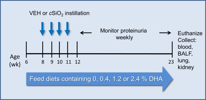
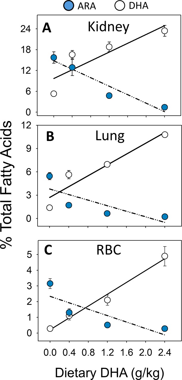
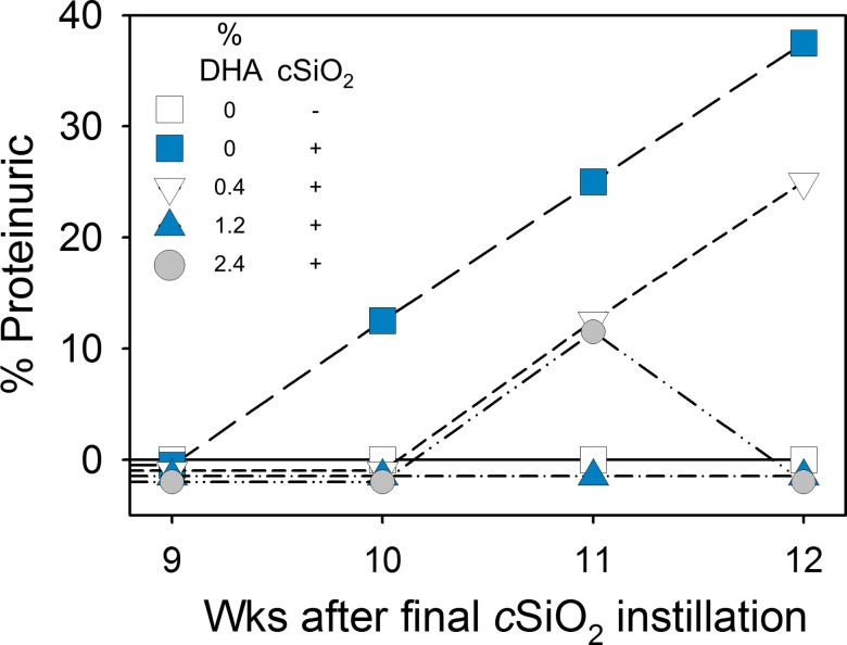
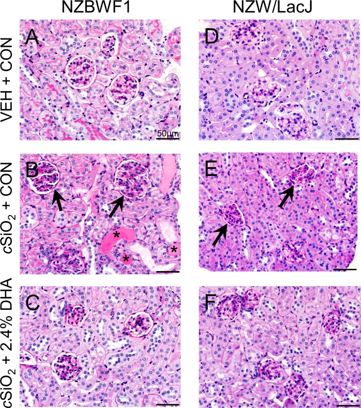
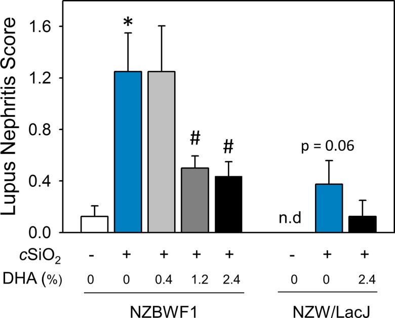
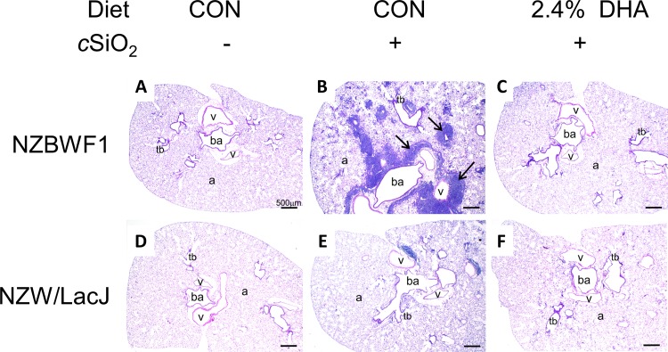
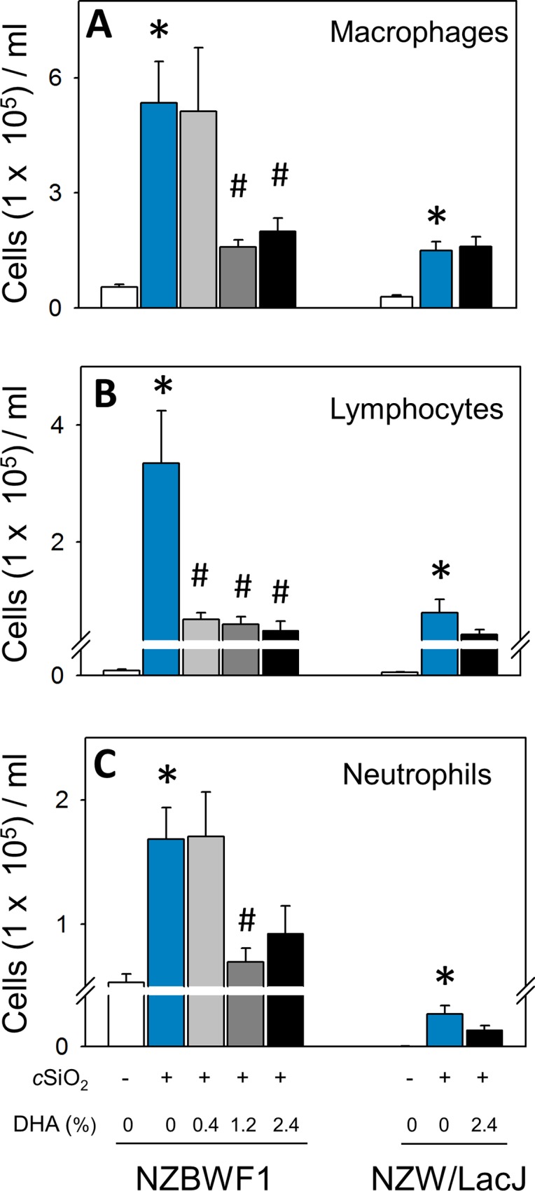
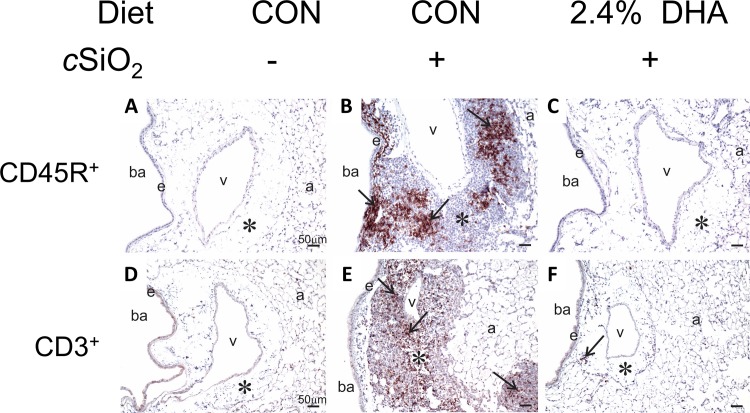
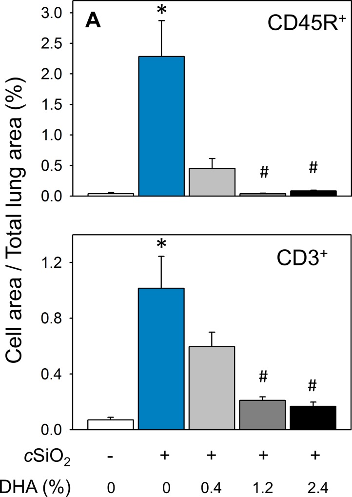
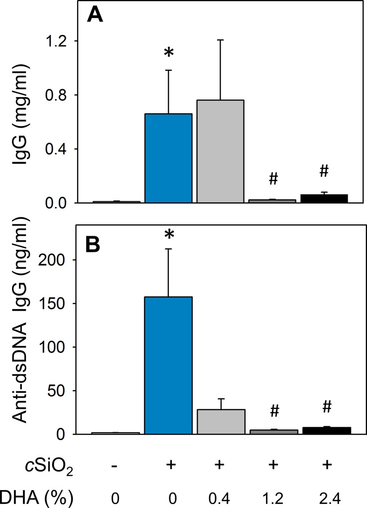
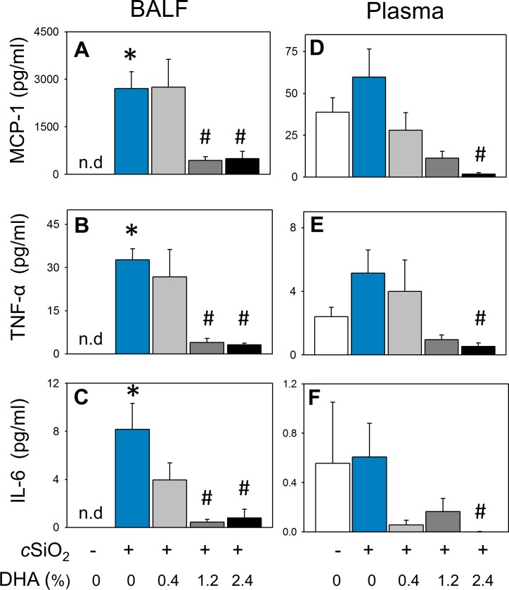
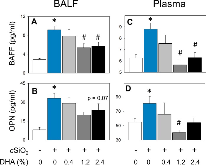
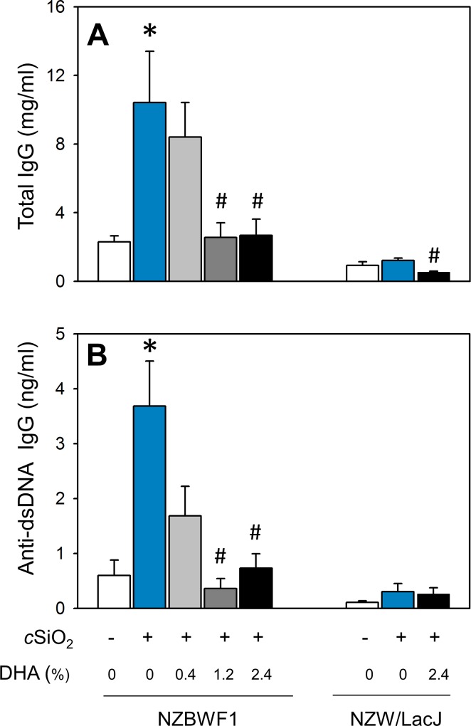
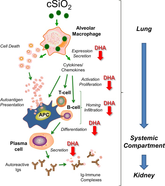
Occupational exposure to respirable crystalline silica (cSiO2, quartz) is etiologically linked to systemic lupus erythematosus (lupus) and other human autoimmune diseases (ADs). In the female NZBWF1 mouse, a widely used animal model that is genetically prone to lupus, short-term repeated intranasal exposure to cSiO2 triggers premature initiation of autoimmune responses in the lungs and kidneys. In contrast to cSiO2's triggering action, consumption of the ω-3 polyunsaturated fatty acid docosahexaenoic acid (DHA) prevents spontaneous onset of autoimmunity in this mouse strain. The aim of this study was to test the hypothesis that consumption of DHA will prevent cSiO2-triggered autoimmunity in the female NZBWF1 mouse. Mice (6 wk old) were fed isocaloric AIN-93G diets containing 0.0, 0.4, 1.2 or 2.4% DHA. Two wk after initiating feeding, mice were intranasally instilled with 1 mg cSiO2 once per wk for 4 wk and maintained on experimental diets for an additional 12 wk. Mice were then sacrificed and the lung, blood and kidney assessed for markers of inflammation and autoimmunity. DHA was incorporated into lung, red blood cells and kidney from diet in a concentration-dependent fashion. Dietary DHA dose-dependently suppressed cSiO2-triggered perivascular leukocyte infiltration and ectopic lymphoid tissue neogenesis in the lung. DHA consumption concurrently inhibited cSiO2-driven elevation of proinflammatory cytokines, B-cell proliferation factors, IgG and anti-dsDNA Ig in both bronchoalveolar lavage fluid and plasma. DHA's prophylactic effects were further mirrored in reduced proteinuria and glomerulonephritis in cSiO2-treated mice. Taken together, these results reveal that DHA consumption suppresses cSiO2 triggering of autoimmunity in female NZBWF1 mice as manifested in the lung, blood and kidney. Our findings provide novel insight into how dietary modulation of the lipidome might be used to prevent or delay triggering of AD by cSiO2. Such knowledge opens the possibility of developing practical, low-cost preventative strategies to reduce the risk of initiating AD and subsequent flaring in cSiO2-exposed individuals. Additional research in this model is required to establish the mechanisms by which DHA suppresses cSiO2-induced autoimmunity and to ascertain unique lipidome signatures predictive of susceptibility to cSiO2-triggered AD.


Conflict of interest statement

Competing Interests: The authors have declared that no competing interests exist.

Figures

Fig 1
Fig 1. Experimental design.
Beginning at age 6 wk, NZBWF1 were assigned CON diet or 0.4, 1.2, and 2.4% DHA diet. NZW/LacJ mice were assigned either CON diet or 2.4% DHA-containing diet. Then, starting at age 8 wk, mice were dosed intranasally with 25 μl PBS VEH or 25 μl PBS containing 1.0 mg cSiO2 weekly for 4 wk. Proteinuria was monitored over the course of the experiment and all animals euthanized at 23 wk of age (i.e. 12 wk PI).
Fig 2
Fig 2. Consumption of microalgal oil dose-dependently increases DHA in kidney, lung and RBC.
DHA incorporation in tissues increased in a dose-dependent manner that coincided with a reduction in ARA. (A) Kidney (r2 = 0.725, P<0.05) (B) lung (r2 = 0.961, p < 0.001), and (C) red blood cell (r2 = 0.876, p < 0.001).
Fig 3
Fig 3. Dietary supplementation with DHA attenuates cSiO2-induced proteinuria in NZBWF1 mice.
Proteinuria (>300 mg/dl) was monitored weekly until sacrifice after the final cSiO2 instillation. VEH-instilled NZBWF1 mice fed CON diet did not develop proteinuria. Proteinuria was undetectable in NZW/LacJ exposed to VEH or cSiO2 over the duration of the experiment.
Fig 4
Fig 4. DHA consumption suppresses cSiO2-induced glomerulonephritis in NZBWF1 mice.
Representative photomicrographs of H&E stained kidney section in NZBWF1 (A-C) and NZW/LacJ (D-F). Letters indicate CON-fed, VEH-exposed mice (A, D), CON-fed, cSiO2-exposed mice (B, E) and 2.4% DHA-fed, cSiO2-exposed mice(C, F). CON-fed NZBWF1 mice instilled with cSiO2 (B) developed extensive glomerulonephritis (black arrows) and tubular proteinosis (*). Mild histopathological lesions were also observed in some NZW/LacJ mice exposed to cSiO2 (E). Dietary supplementation with 2.4% DHA decreased severity of lesions in cSiO2-exposed NZBWF1(C), and NZW/LacJ mice (F).
Fig 5
Fig 5. DHA dose-dependently reduces severity of lupus nephritis in cSiO2-exposed NZBWF1 mice.
NZBWF1 and NZW/LacJ mice were individually graded following the modified ISN/RPS lupus nephritis classification system as described in Materials and Methods. Slide sections from kidneys were graded as follows: (0) no tubular proteinosis; (1) mild tubular proteinosis, early sclerosis, and mild crescent formation; (2) moderate tubular proteinosis, early sclerosis, and crescent formation; (3) marked tubular proteinosis with diffuse global proliferative and sclerosing glomerulonephritis. Data are x¯ ± SEM (n = 8). Symbols: * indicates significant difference from CON-fed mice instilled with VEH (p < 0.05); # indicates significant difference from CON-fed mice instilled with cSiO2 (p < 0.05). DHA dose-dependently reduced cSiO2-triggered lupus nephritis in NZBWF1 mice (r2 = -0.414, p < 0.05).
Fig 6
Fig 6. DHA supplementation prevents cSiO2-induced pneumonitis.
Representative photomicrographs of H&E stained lung sections from NZBWF1 (A-C) and NZW/LacJ (D-F) mice exposed to VEH (A, D), cSiO2 fed CON diet (B, E), and cSiO2 fed 2.4% DHA (C, F). Black arrows in light photomicrographs denote marked leukocyte infiltration that circumvented both the vasculature and airways in the lung following cSiO2 exposure (B). Dietary DHA dramatically reduced cSiO2-induced pulmonary inflammation as evident by the absence of cellular accumulation in (C, F). Lymphocytic cell infiltration was semi-quantitatively graded as indicated in Table 3). Abbreviations: ba = bronchiolar airway, v = blood vessel, tb = terminal bronchiole, a = alveolus.
Fig 7
Fig 7. DHA consumption abrogates cSiO2-induced macrophage, lymphocyte, and polymorphonuclear leukocyte accumulation in BALF.
Differential counts of macrophages (A), lymphocytes (B), and neutrophils (C) in BALF of NZBWF1 and NZW/LacJ mice. Data are x¯ ± SEM (n = 8). Symbols: * indicates significant difference from CON-fed mice instilled with VEH (p < 0.05); # indicates significant difference from CON-fed mice instilled with cSiO2 (p < 0.05). DHA dose-dependently decreased macrophages (r2 = -0.545, p < 0.05), lymphocytes (r2 = -0.599, p < 0.001), and neutrophils (r2 = -0.448, p < 0.05) in NZBWF1 mice.
Fig 8
Fig 8. B and T cell infiltration in lungs of NZBWF1 mice following cSiO2 exposure is prevented by dietary supplementation with DHA.
Representative light photomicrographs of lung tissue sections from CON-fed NZBWF1 mice treated with VEH (A, D), CON-fed NZBWF1 mice treated with cSiO2 (B, E), 2.4% DHA-fed mice treated with cSiO2 (C, F). Lung sections were stained with either CD45R to identify B-lymphocytes (A-C) or CD3 to identify T cells (D-F) and counterstained with hematoxylin. Inflammatory cell infiltrates in peribronchiolar and perivascular interstitium induced by cSiO2 (asterisk in B and E) consisted of both B and T lymphocytes as indicated by positive immunohistochemical staining (black arrows in B and E, respectively). B-lymphocytes tended to form distinct aggregates whereas T lymphocytes, which were more diffusely, scattered throughout lymphoid cells aggregates. Dietary DHA blocked B and T cell accumulation in lungs of cSiO2-treated NZBWF1 mice as evident by marked reduction in CD45R+ and CD3+ cells. Abbreviations: ba = bronchiolar airway, e = airway epithelium, a = alveolus, v = blood vessel, * = interstitium.
Fig 9
Fig 9. cSiO2-triggered B and T cell infiltration in lungs of NZBWF1 mice is dose-dependently prevented by DHA consumption.
Morphometric quantitation of B cell (A) and T cell (B) cellular infiltration in lung parenchyma in CON- and DHA-fed mice exposed to VEH or cSiO2. Data are x¯ ± SEM (n = 8). Symbols: * indicates significant difference from CON-fed mice instilled with VEH (p < 0.05); # indicates significant difference from CON-fed mice instilled with cSiO2 (p < 0.05). DHA consumption dose-dependently decreased CD45R+ (r2 = -0.707, p<0.001) and CD3+ (r2 = -0.728, p<0.001) cellular infiltration.
Fig 10
Fig 10. Dietary DHA suppresses cSiO2 -induced elevation of total IgG and anti-dsDNA Ig in BALF of NZBWF1 mice.
Total IgG (A) and anti-dsDNA Ig (B) in BALF of NZBWF1 mice was quantitated by ELISA. Data are x¯ ± SEM (n = 8). Symbols: * indicates significant difference from CON-fed mice instilled with VEH (p < 0.05); # indicates significant difference from CON-fed mice instilled with cSiO2 (p < 0.05). Dietary DHA dose-dependently decreased total IgG (r2 = -0.574, p < 0.001) and anti-dsDNA Ig (r2 = -0.546, p < 0.05) in BALF.
Fig 11
Fig 11. cSiO2 -induced elevations of proinflammatory cytokines MCP-1, TNF-α and IL-6 in BALF and plasma are decreased by DHA consumption in NZBWF1 mice.
MCP-1 (A, D), TNF-α, (B, E) and IL-6 (C, F)) were quantitated in BALF (left panel) and plasma (right panel) by flow cytometric bead array. Data are x¯ ± SEM (n = 8). The designation n.d. indicates below the limit of detection. Bars without same letter are significantly different (p<0.05). Symbols: * indicates significant difference from CON-fed mice instilled with VEH (p < 0.05); # indicates significant difference from CON-fed mice instilled with cSiO2 (p < 0.05). DHA dose-dependently decreased BALF concentrations of MCP-1 (r2 = -0.791, p < 0.001), TNF- α (r2 = -0.577 p < 0.001), and IL-6 (r2 = -0.810, p < 0.001). DHA dose-dependently decreased plasma MCP-1 (r2 = -0.871, p < 0.001) and TNF-α (r2 = -0.527, p < 0.05).
Fig 12
Fig 12. cSiO2 -induced elevation of B cell stimulating cytokines BAFF and osteopontin are decreased in BALF and plasma in NZBWF1 mice fed DHA.
B cell stimulating cytokines B cell activating factor (BAFF) (A, C) and osteopontin (OPN) (B, D) were quantitated by ELISA in BALF (left panel) and plasma (right panel). Data are x¯ ± SEM (n = 8). Symbols: * indicates significant difference from CON-fed mice instilled with VEH (p < 0.05); # indicates significant difference from CON-fed mice instilled with cSiO2 (p < 0.05). DHA dose-dependently decreased BAFF in BALF (r2 = -0.507, p < 0.05) and plasma (r2 = -0.539, p < 0.05). DHA dose-dependently decreased OPN in BALF (r2 = -0.330, p = 0.06) and in plasma (r2 = -0.493, p < 0.05).
Fig 13
Fig 13. Dietary DHA attenuates cSiO2 -induced elevation of total IgG and anti-dsDNA Ig in plasma of NZBWF1 and NZW/LacJ mice.
Total IgG (A) and anti-dsDNA Ig (B) in plasma of NZBWF1 mice was quantitated by ELISA. Data are x¯ ± SEM (n = 8). Symbols: * indicates significant difference from CON-fed mice instilled with VEH (p < 0.05); # indicates significant difference from CON-fed mice instilled with cSiO2 (p < 0.05). DHA dose-dependently decreased plasma total IgG in NZBWF1 (r2 = -0.493, p < 0.05) and NZW/LacJ mice (r2 = -0.814, p = < 0.001). DHA dose-dependently decreased plasma anti-dsDNA Ig in NZBWF1 (r2 = -0.567, p < 0.001).
Fig 14
Fig 14. Putative mechanisms for DHA-mediated suppression of cSiO2-induced autoimmunity.
The data presented here suggest that cSiO2-triggered pulmonary inflammation and ectopic lymphoid neogenesis drive systemic autoimmunity and glomerulonephritis in the female NZBWF1 mouse. Red downward arrows indicate potential action sites for suppressive effects of DHA that can be further predicted from these data.

References

    1. NIEHS. Autoimmune diseases. 2012. https://www.niehs.nih.gov/health/materials/autoimmune_diseases_508.pdf. Accessed 7/21/2016.
    1. Parks CG, Miller FW, Pollard KM, Selmi C, Germolec D, Joyce K et al. Expert panel workshop consensus statement on the role of the environment in the development of autoimmune disease. Int J Mol Sci. 2014;15(8):14269–14297. 10.3390/ijms150814269 - DOI - PMC - PubMed
    1. OSHA. OSHA's final rule to protect workers from exposure to respirable crystalline silica. 2016. https://www.osha.gov/silica/. Accessed 7/21/2016.
    1. Finckh A, Cooper GS, Chibnik LB, Costenbader KH, Watts J, Pankey H et al. Occupational silica and solvent exposures and risk of systemic lupus erythematosus in urban women. Arthr Rheum. 2006;54(11):3648–3654. 10.1002/art.22210 - DOI - PubMed
    1. Parks CG, Cooper GS, Nylander-French LA, Sanderson WT, Dement JM, Cohen PL et al. Occupational exposure to crystalline silica and risk of systemic lupus erythematosus—A population-based, case-control study in the southeastern United States. Arthr Rheum. 2002;46(7):1840–1850. 10.1002/art.10368 - DOI - PubMed