. 2017 Jul;27(4):480-498.
doi: 10.1111/bpa.12425. Epub 2016 Nov 4.

Long-term survival and regeneration of neuronal and vasculature cells inside the core region after ischemic stroke in adult mice

Affiliations

Long-term survival and regeneration of neuronal and vasculature cells inside the core region after ischemic stroke in adult mice

Michael Qize Jiang et al. Brain Pathol. 2017 Jul.

Abstract

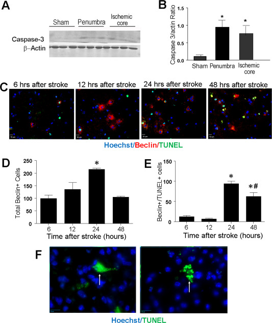
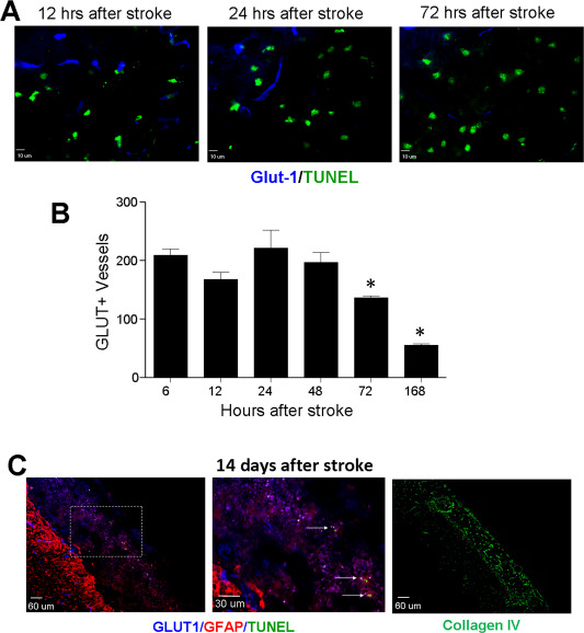
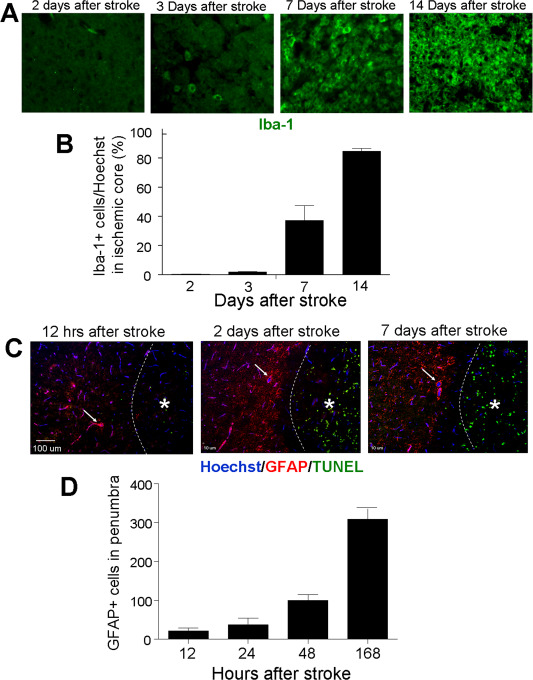
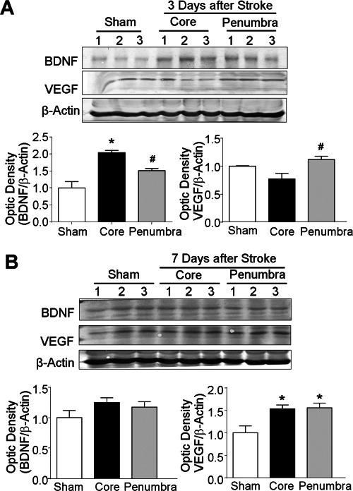
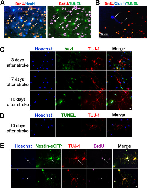
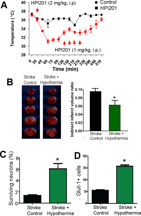
Focal cerebral ischemia results in an ischemic core surrounded by the peri-infarct region (penumbra). Most research attention has been focused on penumbra while the pattern of cell fates inside the ischemic core is poorly defined. In the present investigation, we tested the hypothesis that, inside the ischemic core, some neuronal and vascular cells could survive the initial ischemic insult while regenerative niches might exist many days after stroke in the adult brain. Adult mice were subjected to focal cerebral ischemia induced by permanent occlusion of distal branches of the middle cerebral artery (MCA) plus transient ligations of bilateral common carotid artery (CCA). The ischemic insult uniformly reduced the local cerebral blood flow (LCBF) by 90%. Massive cell death occurred due to multiple mechanisms and a significant infarction was cultivated in the ischemic cortex 24 h later. Nevertheless, normal or even higher levels of brain-derived neurotrophic factor (BDNF) and vascular endothelial growth factor (VEGF) persistently remained in the core tissue, some NeuN-positive and Glut-1/College IV-positive cells with intact ultrastructural features resided in the core 7-14 days post stroke. BrdU-positive but TUNEL-negative neuronal and endothelial cells were detected in the core where extensive extracellular matrix infrastructure developed. Meanwhile, GFAP-positive astrocytes accumulated in the penumbra and Iba-1-positive microglial/macrophages invaded the core several days after stroke. The long term survival of neuronal and vascular cells inside the ischemic core was also seen after a severe ischemic stroke induced by permanent embolic occlusion of the MCA. We demonstrate that a therapeutic intervention of pharmacological hypothermia could save neurons/endothelial cells inside the core. These data suggest that the ischemic core is an actively regulated brain region with residual and newly formed viable neuronal and vascular cells acutely and chronically after at least some types of ischemic strokes.

Keywords: cell survival; ischemic core; neurotrophic factors; neurovasculature; regeneration.


Figures

Figure 1
Figure 1
Focal ischemic stroke in the mouse and ischemia‐induced cell death. Focal cerebral ischemia targeting the right sensorimotor cortex was induced by permanent occlusion of the distal braches of the right MCA and transient (10 minutes) ligation of both CCAs. A. The Laser Doppler scanner was used to measure the local cerebral blood flow (LCBF) in the ischemic region shown on the skull sketch (squire box) on the left. LCBF was reduced to about 10% of basal LCBF during the MCA/CCA blockage. B. 14C‐iodoantipyrine autoradiography was applied to assess regional flow in the brain after MCA/CCA occlusions. The blue area in the right cortex shows a marked and uniformed reduction in LCBF, surrounded by less reduced penumbra. C. One and 3 days after the ischemic insult, TTC staining revealed a well defined ischemic core region in the senserimotor cortex. N = 12 animals for 24 h and 15 for 72 h after stroke. D. DNA damage representing neuronal cell death was identified using TUNEL staining (green) at different times after stroke. In the ischemic core, Hoechst 33342 (blue) was applied to label nuclei of all cells, NeuN (red) was used as a mature neuronal marker. As late as 7 days after stroke, there were still some NeuN+ staining that did not overlay with TUNEL (arrows). E. NeuN+/TUNEL+ cells were counted in the ischemic core region. Quantified cell numbers were from six random survey fields and six brain sections. Neuronal cell death increased from 6 h after stroke and reached to more than 90% at 7 days after stroke. F. Non‐neuronal cells were identified as Hoechst 33342 labeled NeuN negative cells. The number of these cells was relatively stable during the first day after stroke, but showed a reduction at 2 days after stroke. A huge increase of non‐neuronal cells was seen at 7 days after stroke when massive invasion of inflammatory immune cells into the ischemic core. N = 10, Six brain sections were obtained from each animal and six random fields on the core of each section were counted.
Figure 2
Figure 2
Neuronal cell fate in the ischemic core. Immunohistochemical staining was performed in brain sections using NeuN antibody to label neurons and Iba‐1 antibody to label microglial cells in the ischemic cortex. A to D. Neuronal and microglial cells at 7, 14, and 21 days after stroke. NeuN+ cells can be seen inside the ischemic core (*). The enlarged images in C illustrate intact NeuN+ staining (red, arrows) in the core region 14 days after stroke. Meanwhile, there were significant numbers of Iba −1+ migcroglial and macrophages (green) located into the core. Hoechst 33342 staining (blue) marks the nuclei of all cells. E. A magnified image showing Hoechst 33342, NeuN and Iba‐1 labeling in a core area. The NeuN immunoreactivity appears inside the Iba‐1+ cells (arrows), suggesting that neuronal cells and/or debris were cleaned up by microglial/macrophages in the process of phagocytosis. This appears the case for most NeuN positive staining in the core region. F. As a negative control, the image was taken from the core region of a brain section showing the absence of neuron staining with the secondary Cy3 antibody but without adding the primary antibody NeuN.
Figure 3
Figure 3
Cell death and survival in the ischemic core after embolic stroke of sever ischemia. A severe ischemic stroke was induced in adult mice by permanent occlusion of the MCA using an autologous blood clot. A. TTC staining of brain sections at 1 and 4 days after the ischemic insult. B. Immunohistochemical staining of NeuN (red), TUNEL (green) and Hoechst 33342 (blue) in the core region for the inspection of neuronal cell death at 7 days after stroke. NeuN positive but TUNEL negative cells can be seen in the region. The frames show the enlarged areas in C and D. C. Three dimensional images show a NeuN positive but TUNEL negative neurons (arrow). D. Glut‐1 staining was used for vascular endothelial cells. The 3‐D images show endothelial cells that were TUNEL negative. Representative of three animals.
Figure 4
Figure 4
Apoptosis and autophagy contributed to cell death in the ischemic core. Signals in the apoptotic and autophagic cascades were examined in the ischemic cortex. A and B. Western blotting shows activation of caspase‐3 in the penumbra and core regions 1 days after stroke. N = 7. *. P < 0.05 vs. sham controls. C. The autophagy marker beclin 1 was detected in immunostaining of the ischemic core area (arrows). D. Quantification of total beclin 1+ cells per animal (six brain sections and six random fields on each section) in the ischemic core different time points after stroke. E. Double labeling of beclin 1 and TUNEL revealed a relationship between the beclin 1 immunoreactivity and cell death, especially 24 to 48 h after stroke. Comparing to the bar graph in D, approximately 50% of beclin cells were also TUNEL+ at these time points. N = 3 animals per time point. *. P < 0.05 vs. 6 h, #. P < 0.05 vs. 24 h. F. Differential morphology of TUNEL+ neurons. The image on the left shows smear TUNEL staining without noticeable nucleus fragmentation (Type 1 TUNEL+ cells; arrow). The image on the right shows fragmented DNA of TUNEL staining (Type 2 TUNEL+ cells; arrow).
Figure 5
Figure 5
Vascular endothelial cells and microvessel structures in the ischemic core. Vascular endothelial cells were examined using the glucose transporter 1 antibody in immunohistochemical examination. A. Immunoflourenscent images of Glut‐1 (blue) and TUNEL (green) staining in the ischemic core at different days after stroke. B. Quantified data of the experiment in A. The number of Glut‐1+ endothelial cells/vessels remained relatively constant for up to 2 days after ischemia. Glut‐1+ cells decreased gradually from 3 to 7 days after stroke. N = 7 per time point, *. P < 0.05 vs. 6 h data). C. At 14 days after stroke, Glut‐1 (blue) markers was still widely detectable inside the ischemic core, while GFAP labeled glial cells (astrocytes) were mostly located in the peri‐infarct region. In the enlarged image from the frame shown in the left, TUNEL staining revealed some cell death process at this very delayed time point. The Collagen IV staining verified the extramatrix networks developed in the ischemic core 14 days after stroke.
Figure 6
Figure 6
Distribution of microglia/macrophages and astrocytes in the ischemic and penumbra regions after stroke. Immunohistochemical staining was performed to track the invasion and localization of Iba‐1+ and GRAP+ cells in the ischemic cortex. A and B. Iba‐1 staining (green) in the ischemic core at different days after stroke. Iba‐1+ microglial/macrophages were rare in the core during the first few days after stroke. These cells started to occupy the ischemic core tissue several days after stroke, by day 14 about 90% of cells in the core were Iba‐1+ cells. N = 10 animals. Six brain sections were obtained from each animal and six random fields on the core of each section were counted. C and D. GFAP staining (red) was applied to identify activated astrocytes and TUNEL staining (green) was used to detect dead cells. Hoechst 33342 (blue) labeled nuclei of all cells. GFAP+ astrocytes started to accumulate in the peri‐infarct area 12 h after stroke, and the number increased gradually as long as 7 days after stroke. Arrows point to some GFAP/Hoechst positive cells in the peri‐infarct area. TUNEL staining was concentrated in the ischemic core (*), demonstrating massive cell death in the core. The bar graph shows the average total counted numbers of cells per animal. N = 8–12 animals. Six brain sections were obtained from each animal and six random fields on the core of each section were counted.
Figure 7
Figure 7
Ultrastructural evidence of surviving cells in the ischemic core 7 days after stroke. A transmission electron microscope was used to examine the control brain section and the ischemic core of brain sections 7 days after ischemia. A. A control EM image of normal cortical neurons in the contra lateral cortex. The neurons had clear boundaries and a large nuclear (*), chromatin was uniformly distributed, the plasma membrane was continuous and clear, and many organelles including mitochondria and endoplasmic reticulum (ER) were visible. B. A representative surviving neuronal cells in the ischemic core. The cell contains a relatively large and universal nucleus, although the total cell size appeared smaller than the control neurons. The cell was surrounded by an intact plasma membrane and abundant cellular organelles existed in the cytoplasm. C. Another example of surviving neuronal cells in the ischemic core. In addition to the near normal nucleus (*) and chromatin, there were numerous intracellular organelles including mitochondria and ER (arrows) contained in the axonal shape of the cell and the intact membrane. Some lysosomes existed in the cytoplasm, suggesting the progress of degeneration. D. Remaining synaptic structures in the ischemic core region. Several presynaptic terminals and transmitter vesicles (V) inside the terminal can be seen in this image. The dark post‐synaptic density (PSD) was located opposite to the pre‐synaptic membrane. E. A microglial or microphagic cell showing two processes containing many lysosomes in the cytosol. Note the small size of this type of cell. F. Myelinated axon as shown in this image can be seen in the ischemic core. The image shows a cross section of the myelin (M) surrounding nerve fibers. G. Some autophagosomes (A) were identified, suggesting the process of autophagy. H and I. Surviving microvessels and endothelial cells (E) as well as the infrastructure of the neurovascular unit were easily detectable in the ischemic core. Astrocytes and/or pericytes (#) and basement membrane were surrounding endothelial cells. The lumen space was clearly formed inside the neurovascular unit.
Figure 8
Figure 8
Expression of trophic factors in the ischemic core and penumbra. Western blotting was applied to assay the expression levels of BDNF and VEGF in the ischemic core and penumbra regions 3 and 7 days after stroke. A. BDNF and VEGF expression levels 3 days after stroke in sham control, core and penumbra brain tissues. B. BDNF and VEGF levels at 7 days after stroke in the three brain regions. The bar graph illustrates the expression ratios normalized to sham control after correction with loading controls. *. P < 0.05 vs. sham, #. P < 0.05 vs. core; n = 6 animals per group.
Figure 9
Figure 9
Regenerative niche exited in the ischemic tissue. Regenerative activities in the ischemic core were inspected in the brain and under cultured conditions. A and B. Stroke animals received BrdU injections daily to label proliferating cells. Seven days after stroke, immunostaining of the core region revealed the existence of BrdU/NeuN double positive cells that were TUNEL negative (A). BrdU/Glut1 positive but TUNEL negative endothelial cells were also observed in the core (B). Arrows point to BrdU/NeuN or BrdU/Glut1 double positive cells. To detect regenerative niche that might reside in the ischemic core tissue, we dissected the core tissue at 7 days after stroke and plated the dissociated cells in PDL/Laminin coated dishes. C. Different days after in vitro, cells were fixed and stained with immature neuronal marker TUJ‐1 (red) and microglia cell marker Iba‐1 (green). There were increasing numbers of TUJ‐1+ cells from 3 to 10 days after stroke. Many of them did not overlay with Iba1. D. In these images taken 10 days after stroke, TUJ‐1+ cells (red) were negative for TUNEL (green) staining suggesting they were viable cells. Scale bar = 10 μm. E. Nestin‐eGFP transgenic mice were subjected to the focal ischemic stroke. BrdU (50 mg/kg) was injected daily from 1 day after stroke. Cells from the ischemic core of 7 days after stroke were cultured for 7 days. Nestin‐eGFP (green) was easily detectable in this culture and it overlaid with the immature neuronal marker TUJ‐1 (red) as well as with the proliferation marker BrdU (purple) (arrow). There were a few cells that were eGFP and TUJ‐1 positive but BrdU negative (arrowhead), implying that they might be surviving original cells. Scale bar = 10 μm.
Figure 10
Figure 10
Protection of the neuronal and vascular cells in the ischemic core. A. The neurotensin receptor 1 agonist HPI‐201 was injected 60 minutes after the onset of ischemia. HPI‐201 injections effectively reduced the body temperature of the stroke animals from 37°C to 32°C for 6 h. B. TTC staining of the ischemic brain sections 3 days after focal ischemia. HPI‐201‐treated animals showed significantly reduced infarct volume compared to stroke/saline controls. N = 5 per group, *. P < 0.05 vs. stroke control. C and D. HPI‐201 treatment significantly increased surviving NeuN+ neurons (C) and endothelial cells (D) in the core 7 days after stroke. N = 7, *. P < 0.05 vs. stroke controls.

Similar articles

Cited by

References

    1. Aronowski J, Cho KH, Strong R, Grotta JC (1999) Neurofilament proteolysis after focal ischemia; when do cells die after experimental stroke? J Cereb Blood Flow Metab 19:652–660. - PubMed
    1. Back T (1998) Pathophysiology of the ischemic penumbra–revision of a concept. Cell Mol Neurobiol 18:621–638. - PMC - PubMed
    1. Barber PA, Davis SM, Infeld B, Baird AE, Donnan GA, Jolley D, Lichtenstein M (1998) Spontaneous reperfusion after ischemic stroke is associated with improved outcome. Stroke 29:2522–2528. - PubMed
    1. Baron JC, Yamauchi H, Fujioka M, Endres M (2014) Selective neuronal loss in ischemic stroke and cerebrovascular disease. J Cereb Blood Flow Metab 34:2–18. - PMC - PubMed
    1. Barone FC (2009) Ischemic stroke intervention requires mixed cellular protection of the penumbra. Curr Opin Investig Drugs 10:220–223. - PubMed

Publication types

MeSH terms