. 2016 Aug 11;15(1):127.
doi: 10.1186/s12944-016-0294-4.

Lipopolysaccharide induces SBD-1 expression via the P38 MAPK signaling pathway in ovine oviduct epithelial cells

Affiliations

Lipopolysaccharide induces SBD-1 expression via the P38 MAPK signaling pathway in ovine oviduct epithelial cells

Qi Li et al. Lipids Health Dis. .

Abstract

Background: Beta defensins are secreted from ovine oviduct epithelial cells (OOECs) in response to microbial infection, and are potential alternatives to antibiotic agents in the treatment of microorganism infection, particularly given the abuse of antibiotic agents and the increasing number of drug-resistant bacteria. The aberrant expression of defensins may result in disorders involving organ and oviduct inflammation, such as salpingitis.

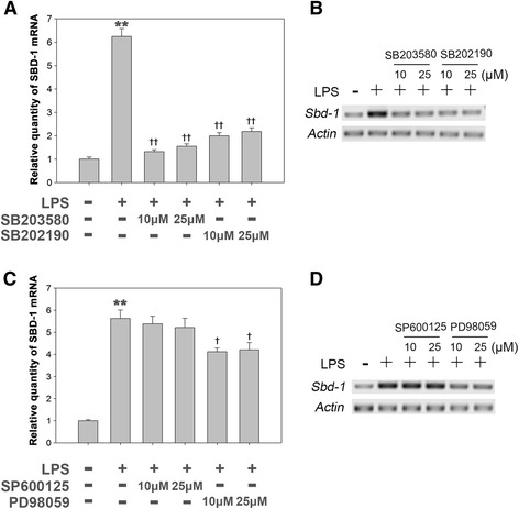
Methods: In the present study, we investigated the effects of LPS on the mRNA expression levels of sheep β-defensin-1 (SBD-1) in ovine oviduct epithelial cells. The OOECs in vitro culturing system were established and treated with different concentrations of LPS for indicated time. In addition, MAPK inhibitors and TLR4 antibodies were pretreated to investigate the potential mechanism which involves in LPS regulating SBD-1 expression.

Results: LPS markedly upregulated SBD-1 expression in a concentration- and time-dependent manner. Treatment with 100 ng/mL LPS resulted in the phosphorylation of JNK, ERK and P38 MAPK. Interestingly, the LPS stimulated SBD-1 expression was attenuated by pretreatment with the P38 MAPK inhibitors SB203580 and SB202190 but not the JNK inhibitor SP600125, while the ERK inhibitor PD98059 had a minor effect. Furthermore, treatment with a Toll-like receptor 4 (TLR4) neutralizing antibody significantly decreased P38 MAPK phosphorylation and LPS induced SBD-1 expression.

Conclusions: Together, these findings suggest that SBD-1 is upregulated by LPS via the TLR4 receptor, mainly through the P38 MAPK signaling pathway in ovine oviduct epithelial cells to protect the ovine oviduct epithelium from pathogen invasion.

Keywords: LPS; Ovine oviduct epithelial cells; P38 MAPK; SBD-1; TLR4.


Figures

Fig. 1
Fig. 1
LPS induces SBD-1 mRNA expression and activates the MAPK signaling pathway in ovine oviduct epithelial cells. a Immunofluorescence was performed to identify ovine oviduct epithelial cells. After 24 h in culture, cells were stained for an ovine oviduct epithelial cell marker CK-18 (green). Hoechst 33342 (blue) was used to counterstain the nuclear DNA. An isotype-matched IgG was used as the negative control. Scale bar: 100 μm. b Ovine oviduct epithelial cells (OOECs) were treated with the indicated concentrations of LPS for 24 h and compared with untreated controls. The QRT-PCR analysis showed that LPS induces SBD-1 expression in a concentration-dependent manner. c OOECs were treated with LPS (100 ng/mL) for various time intervals and compared with untreated controls. The QRT-PCR results indicated that LPS induces SBD-1 expression in a time-dependent manner. d OOECs were treated with LPS (100 ng/mL) and harvested at the indicated time points. Whole-cell lysates were prepared and used for western blot analysis with MAPK and P-MAPK antibodies. e A densitometric analysis of the optical density (OD) of different p-MAPKs relative to the OD of MAPKs. All of the experiments were repeated at least three times. *p < 0.05 vs control (t-test)
Fig. 2
Fig. 2
LPS induces SBD-1 expression mainly through P38 MAPK signaling but not through JNK or ERK signaling. a b OOECs were cultured with LPS (100 ng/mL), with or without P38 MAPK inhibitors SB203580 and SB202190 for 12 h. Total RNA was prepared and used for examination of SBD-1 mRNA expression by QRT-PCR or RT-PCR with specific primers for SBD-1 and GAPDH. c d OOECs were cultured with LPS (100 ng/mL), with or without JNK inhibitor SP600125 and ERK inhibitor PD98059 for 12 h. Total RNA was prepared and used for examination of SBD-1 mRNA expression by QRT-PCR or RT-PCR with specific primers for SBD-1 and GAPDH. All of the experiments were repeated at least three times. *p < 0.05, **p < 0.01 (t-test) vs. control. †p < 0.05, ††p < 0.01 (t-test) vs. LPS
Fig. 3
Fig. 3
The cellular localization of P38 MAPK in ovine oviducts. a Immunohistochemistry was performed to determine the cellular localization of P38 and P-P38 in the ovine oviduct. P38 was detected in the epithelial cells but not the stromal cells in the ovine oviduct. The expression pattern of P-P38 was consistent with that of P38. Ovine oviducts were immunohistochemically stained with rabbit IgG as a negative control. Scale bars: 20 μm. b Western blotting experiments were conducted to examine the P38 and P-P38 expression in ovine oviducts in order to detect the specificity of these antibodies
Fig. 4
Fig. 4
LPS stimulates the expression of SBD-1 through TLR4. a Tlr4 mRNA expression of ovine oviduct stromal cells (OOSCs) and epithelial cells (OOECs) was assessed by qRT-PCR. b PCR was conducted to examine the expression of TLR4 in the ovine oviduct epithelial cells harvested at different times. c Western blotting was performed to examine the expression levels of P38 MAPK after treatment with LPS (100 ng/mL) or a TLR4 neutralizing antibody for 12 h. d The densitometric analysis of the bands on the western blotting showed that LPS could markedly activate P38 MAPK, while the separate addition of the TLR4 neutralizing antibody had no effect. However, treatment with the TLR4 neutralizing antibody could significantly decrease the levels of phosphorylated P38 induced by LPS. e QRT-PCR analysis was used to examine the mRNA levels of SBD-1 after treatment with the TLR4 neutralizing antibody for 12 h. Blocking TLR4 activity could inhibit the expression of SBD-1. All of the experiments were repeated at least three times. *p < 0.05, **p < 0.01 (t-test) vs. Control. †p < 0.05, ††p < 0.01 (t-test) vs. LPS
Fig. 5
Fig. 5
Schematic diagram of LPS regulating SBD-1 expression. LPS binds to TLR4 receptor, activates P38 MAPK signaling pathway, and subsequently stimulates SBD-1 expression in ovine oviduct epithelial cells

References

    1. Cohen CR, Mugo NR, Astete SG, Odondo R, Manhart LE, et al. Detection of Mycoplasma genitalium in women with laparoscopically diagnosed acute salpingitis. Sex Transm Infect. 2005;81:463–6. doi: 10.1136/sti.2005.015701. - DOI - PMC - PubMed
    1. Tohidnezhad M, Varoga D, Podschun R, Wruck CJ, Seekamp A, et al. Thrombocytes are effectors of the innate immune system releasing human beta defensin-3. Injury. 2011;42:682–6. doi: 10.1016/j.injury.2010.12.010. - DOI - PubMed
    1. Diamond G, Beckloff N, Ryan LK. Host defense peptides in the oral cavity and the lung: similarities and differences. J Dent Res. 2008;87:915–27. doi: 10.1177/154405910808701011. - DOI - PMC - PubMed
    1. Rehaume LM, Hancock RE. Neutrophil-derived defensins as modulators of innate immune function. Crit Rev Immunol. 2008;28:185–200. doi: 10.1615/CritRevImmunol.v28.i3.10. - DOI - PubMed
    1. Ganz T. Defensins: antimicrobial peptides of innate immunity. Nat Rev Immunol. 2003;3:710–20. doi: 10.1038/nri1180. - DOI - PubMed

MeSH terms