. 2016 Oct 1;311(4):G634-G647.
doi: 10.1152/ajpgi.00092.2016. Epub 2016 Aug 11.

Soluble bioactive microbial mediators regulate proteasomal degradation and autophagy to protect against inflammation-induced stress

Affiliations

Soluble bioactive microbial mediators regulate proteasomal degradation and autophagy to protect against inflammation-induced stress

Yuhei Inaba et al. Am J Physiol Gastrointest Liver Physiol. .

Abstract

Bifidobacterium breve and other Gram-positive gut commensal microbes protect the gastrointestinal epithelium against inflammation-induced stress. However, the mechanisms whereby these bacteria accomplish this protection are poorly understood. In this study, we examined soluble factors derived from Bifidobacterium breve and their impact on the two major protein degradation systems within intestinal epithelial cells, proteasomes and autophagy. Conditioned media from gastrointestinal Gram-positive, but not Gram-negative, bacteria activated autophagy and increased expression of the autophagy proteins Atg5 and Atg7 along with the stress response protein heat shock protein 27. Specific examination of media conditioned by the Gram-positive bacterium Bifidobacterium breve (Bb-CM) showed that this microbe produces small molecules (<3 kDa) that increase expression of the autophagy proteins Atg5 and Atg7, activate autophagy, and inhibit proteasomal enzyme activity. Upregulation of autophagy by Bb-CM was mediated through MAP kinase signaling. In vitro studies using C2BBe1 cells silenced for Atg7 and in vivo studies using mice conditionally deficient in intestinal epithelial cell Atg7 showed that Bb-CM-induced cytoprotection is dependent on autophagy. Therefore, this work demonstrates that Gram-positive bacteria modify protein degradation programs within intestinal epithelial cells to promote their survival during stress. It also reveals the therapeutic potential of soluble molecules produced by these microbes for prevention and treatment of gastrointestinal disease.

Keywords: Atg5; Atg7; cell death; cell survival; host-microbe interaction; inflammatory bowel diseases; microbiota; microflora; mucosal inflammation; peptidoglycan.


Figures

Fig. 1.
Fig. 1.
Conditioned media (CM) from representative Gram-positive, but not Gram-negative, microbes increase expression of heat shock protein 27 (Hsp27) and autophagy proteins while also activating autophagy in C2BBe1 cells. A: CM from representative Gram-positive bacteria activate autophagy and increase Hsp27 expression. C2BBe1 cells were treated with 1% CM from selected Gram-positive [Bifidobacterium breve (BB), Lactobacillus rhamnosus GG (LGG), and Lactobacillus plantarum (LP)] and Gram-negative [Enterococcus faecalis (EF), Proteus mirabilis (PM), and Pseudomonas aeruginosa (PA)] bacteria for 8 h, and proteins were analyzed by Western blotting. Images shown are representative of 3 separate experiments. B: CM from Bifidobacterium breve (Bb-CM) induces autophagy in C2BBe1 cells. Cells were treated with 1% Bb-CM for 8 h and fixed, and autophagosomes were visualized using anti-LC3 antibody (green). DAPI was included during fixation to stain nuclei (blue). Accumulation of LC3 in autophagosomes was quantified using ImageJ software. C: Bb-CM increases Atg5 and Atg7 and decreases p62 in C2BBe1 cells in a concentration- and time-dependent manner. Cells were treated with varying concentrations for 8 h or for varying times with 1% CM, and proteins were extracted and analyzed by Western blotting. Values are means ± SE for 3 separate experiments for A and C, and for B at least 300 cells were counted in each experiment. *P < 0.05, +P < 0.01, ++P < 0.001.
Fig. 2.
Fig. 2.
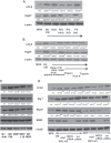
Bb-CM stimulates multiple MAP kinase pathways and increases expression of a number of autophagy proteins. A: p38 activity is increased by CM from Gram-positive bacteria. C2BBe1 were incubated with 1% CM for 8 h from various bacteria as in Fig. 1A. B: Bb-CM activates p38, JNK, and ERK1/2 signaling. C2BBe1 were treated with Bb-CM for the indicated times and at the indicated concentrations. C: Bb-CM acts through MAPK pathways to activate autophagy and increase Hsp27 expression. Cells were treated with specific inhibitors of the MAP kinase pathways for 2 h before 1% Bb-CM for 8 h, and proteins were analyzed by Western blotting. SB, p38 inhibitor SB-203580; SP, JNK inhibitor SP-600125; PD, ERK inhibitor PD-98059; LY, Akt inhibitor LY-294002. Densitometric density was measured using ImageJ software. The no treatment control was set to 100, and values presented are means ± SE for 4 separate experiments. *P < 0.05, +P < 0.01, ++P < 0.001 compared with zero time MRS control by ANOVA.
Fig. 3.
Fig. 3.
Small molecular mass compounds of Bb-CM activate autophagy, whereas small and large molecules induce Hsp27. A: Bb-CM activity is found in the 3-kDa filtrate fraction. Bb-CM was treated with heat or size fractionated using 10- and 3-kDa cutoff filters and then used to treat C2BBe1 cells (1% vol/vol dilution for 8 h). B: Bb-CM activity is altered by protease treatment. Bb-CM was size fractionated (10-kDa cutoff filter) and treated with the proteases pepsin, proteinase K, and trypsin and then used to treat C2BBe1 cells (8 h at 1% vol/vol dilution). 10-kDa cutoff filters were used after digestion to remove protease (all proteases used at 50 μg/ml). C: N-acetylmuramyl-l-alanyl-d-isoglutamine (MDP) treatment does not replicate Bb-CM administration. Unmodified MDP (1 μg/ml), lysine-18 sterol conjugated (l-18 MDP, 100 ng/ml), or the inactive DD amino acid MDP isomer (DD MDP 10 μg/ml) were administered at 10 μg/ml for 8 h of treatment, and protein expression was determined by Western blotting. D: blocking MDP sensing does not block Bb-CM activity. C2BBe1 cells were treated with inhibitor gefitinib, or NOD2 was silenced using siRNA and a control siRNA, which does not recognize any mouse target used (from Ambion). The cells were then treated with Bb-CM or MDP. Images in all panels are representative of 3 separate experiments. Densitometry was performed using ImageJ. The unstimulated control was set to zero, and values are presented are means ± SE. *P < 0.05, +P < 0.01, ++P < 0.001 compared with unstimulated control by ANOVA.
Fig. 4.
Fig. 4.
Bb-CM contains small molecular mass, acetonitrile-extractable factors that inhibit proteasome activity. A: Bb-CM blocks proteasomal enzyme activity. C2BBe1 cells were treated for 8 h with 1% Bb-CM or size fractionated, heat treated, proteinase K treated, or treated with acetonitrile extractable factors. The lysosomal inhibitor bafilomycin A or the combination of proteasome inhibitors MG132 and lactacystin were also used to treat the cells. The 3 proteolytic activities of the proteasome were then measured using specific fluorescence-labeled substrates. Values at 60 min are presented in the bottom, right, and statistical difference from untreated control was determined by ANOVA using a Bonferroni correction. B: proteasomal inhibition correlates with autophagy induction by Bb-CM. Proteins from cells treated in A were analyzed by Western blotting for designated autophagy proteins and Hsp27. Data presented are representative and from 1 experiment repeated 4 times. Densitometric values were determined using ImageJ, control was set to 100%, and statistical difference was determined by ANOVA, *P < 0.05, +P < 0.01.
Fig. 5.
Fig. 5.
Silencing Atg7 increases apoptosis stimulated by hydrogen peroxide but not TNF-α or FAS receptor activation. C2BBe1 cells were transfected with a silencing oligonucleotide for Atg7 or a scrambled oligonucleotide and then treated with IFN-γ (to increase TNF-α receptor expression) or IL-2 (to increase FAS receptor expression) and then Bb-CM. 8 h after Bb-CM, cells were stimulated with hydrogen peroxide (100 μM) (A), TNF-α (100 ng/ml) (B), or anti-Fas receptor (CH-11, 10 μg/ml) (C). Cells were harvested 4 h later and analyzed by Western blotting. Images shown are representative of 3 separate experiments. PARP, poly(ADP-ribose) polymerase.
Fig. 6.
Fig. 6.
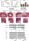
Epithelial cell deletion of Atg7 sensitizes mice to dextran sodium sulfate (DSS)-induced colitis. A mouse line with floxed Atg7 alleles was bred to villin-Cre mice, and littermates with both Atg7 floxed alleles with and without Cre driven by the villin promoter were identified by genotyping. Mice of each type were treated for 7 days with 100 μl Bb-CM or MRS broth by oral gavage before DSS administration (5 days). Mice were gavaged daily during DSS treatment with Bb-CM or MRS as appropriate. Body weight (A) and disease activity index (B) were determined each day. Distal colon damage (C) was scored in histological sections by a blinded reviewer, and cytokine levels were measured (D) in mice killed at day 5 (n = 4 for each group). Mice (n = 3 for each group) were killed, and colonic protein homogenates were analyzed by Western blotting for caspase-3 and PARP (E) as well as LC3, p62, and Atg7 (F). For body weight, disease activity index, and cytokines, analysis was performed by Kruskal-Wallis test with a Dunn's multiple comparison. For body weight and disease activity, significance values (*P < 0.05, +P < 0.01, ++P < 0.001) were always compared with condition of least injury (without Cre, with Bb-CM). For cytokines the comparison is to the highest value, with Cre and no Bb-CM.

References

    1. Adolph TE, Tomczak MF, Niederreiter L, Ko HJ, Bock J, Martinez-Naves E, Glickman JN, Tscurtschenthaler M, Hartwig J, Hosomi S, Fiak MB, Cusick JL, Kohno K, Iwawaki T, Billman-Born S, Raine T, Bharti R, Lucius R, Kweon MN, Marciniak SJ, Choi A, Hagen SJ, Schreiber S, Rosensteil P, Kaser A, Blumberg RS. Paneth cells as a site of origin for intestinal inflammation. Nature 503: 272–276, 2013. - PMC - PubMed
    1. Atarashi K, Tanoue T, Oshima K, Suda W, Nagano Y, Nishikawa H, Fukuda S, Saito T, Narushima S, Hase K, Kim S, Fritz JV, Wilmes P, Ueha S, Matsushima K, Ohno H, Olle B, Sakaguchi S, Taniguchi T, Morita H, Hattori M, Honda K. Treg induction by a rationally selected mixture of Clostridia strains from the human microbiota. Nature 500: 232–236, 2013. - PubMed
    1. Basler M, Dajee M, Moll C, Groettrup M, Kirk CJ. Prevention of experimental colitis by a selective inhibitor of the immunoproteasome. J Immunol 185: 634–641, 2010. - PubMed
    1. Benbrook DM, Long A. Integration of autophagy, proteasomal degradation, unfolded protein response and apoptosis. Exp Oncol 34: 286–297, 2012. - PubMed
    1. Bertolotti A, Wang X, Novao I, Jungreis R, Schlessinger K, Cho JH, West AB, Ron D. Increased sensitivity to dextran sulfate sodium in IRE1beta deficient mice. J Clin Invest 107: 585–593, 2001. - PMC - PubMed

Publication types

MeSH terms

Substances