

Heterologous expression of antigenic peptides in Bacillus subtilis biofilms
- PMID: 27514610
- PMCID: PMC4982213
- DOI: 10.1186/s12934-016-0532-5
Heterologous expression of antigenic peptides in Bacillus subtilis biofilms
Abstract
Background: Numerous strategies have been developed for the display of heterologous proteins in the surface of live bacterial carriers, which can be used as vaccines, immune-modulators, cancer therapy or bioremediation. Bacterial biofilms have emerged as an interesting approach for the expression of proteins of interest. Bacillus subtilis is a well-described, endospore-forming organism that is able to form biofilms and also used as a probiotic, thus making it a suitable candidate for the display of heterologous proteins within the biofilm. Here, we describe the use of TasA, an important structural component of the biofilms formed by B. subtilis, as a genetic tool for the display of heterologous proteins.
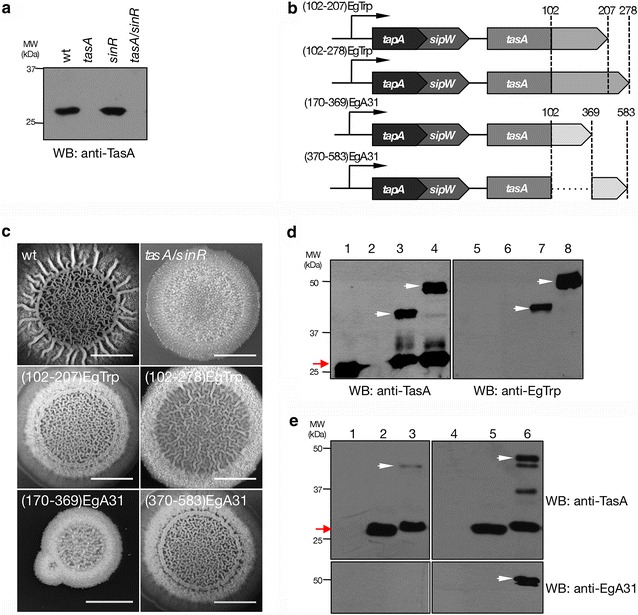
Results: We first engineered the fusion protein TasA-mCherry and showed that was widely deployed within the B. subtilis biofilms. A significant enhancement of the expression of TasA-mCherry within the biofilm was obtained when depleting both tasA and sinR genes. We subsequently engineered fusion proteins of TasA to antigenic peptides of the E. granulosus parasite, paramyosin and tropomyosin. Our results show that the antigens were well expressed within the biofilm as denoted by macrostructure complementation and by the detection of the fusion protein in both immunoblot and immunohistochemistry. In addition, we show that the recombinant endospores of B. subtilis preserve their biophysical and morphological properties.
Conclusions: In this work we provide strong evidence pointing that TasA is a suitable candidate for the display of heterologous peptides, such as antigens, cytokines, enzymes or antibodies, in the B. subtilis biofilms. Finally, our data portray that the recombinant endospores preserve their morphological and biophysical properties and could be an excellent tool to facilitate the transport and the administration.
Keywords: Antigen; Bacillus subtilis; Biofilm; E. granulosus; Endospores; Heterologous protein; Paramyosin; TasA; Tropomyosin; mCherry.
Figures


Similar articles
-
Oral Application of Recombinant Bacillus subtilis Spores to Dogs Results in a Humoral Response against Specific Echinococcus granulosus Paramyosin and Tropomyosin Antigens.Infect Immun. 2018 Feb 20;86(3):e00495-17. doi: 10.1128/IAI.00495-17. Print 2018 Mar. Infect Immun. 2018. PMID: 29229735 Free PMC article.
-
Display of Heterologous Proteins in Bacillus Subtilis Biofilms for Enteric Immunization.Methods Mol Biol. 2022;2465:73-95. doi: 10.1007/978-1-0716-2168-4_4. Methods Mol Biol. 2022. PMID: 35118616
-
Mouse intestinal microbiota reduction favors local intestinal immunity triggered by antigens displayed in Bacillus subtilis biofilm.Microb Cell Fact. 2018 Nov 26;17(1):187. doi: 10.1186/s12934-018-1030-8. Microb Cell Fact. 2018. PMID: 30477481 Free PMC article.
-
Exploitation of Bacillus subtilis as a robust workhorse for production of heterologous proteins and beyond.World J Microbiol Biotechnol. 2018 Sep 10;34(10):145. doi: 10.1007/s11274-018-2531-7. World J Microbiol Biotechnol. 2018. PMID: 30203131 Review.
-
Recent progress in Bacillus subtilis spore-surface display: concept, progress, and future.Appl Microbiol Biotechnol. 2017 Feb;101(3):933-949. doi: 10.1007/s00253-016-8080-9. Epub 2017 Jan 6. Appl Microbiol Biotechnol. 2017. PMID: 28062973 Review.
Cited by
-
Progress in research and application development of surface display technology using Bacillus subtilis spores.Appl Microbiol Biotechnol. 2020 Mar;104(6):2319-2331. doi: 10.1007/s00253-020-10348-x. Epub 2020 Jan 27. Appl Microbiol Biotechnol. 2020. PMID: 31989224 Free PMC article. Review.
-
Oral Application of Recombinant Bacillus subtilis Spores to Dogs Results in a Humoral Response against Specific Echinococcus granulosus Paramyosin and Tropomyosin Antigens.Infect Immun. 2018 Feb 20;86(3):e00495-17. doi: 10.1128/IAI.00495-17. Print 2018 Mar. Infect Immun. 2018. PMID: 29229735 Free PMC article.
-
Hydrodynamic Effects on Biofilm Development and Recombinant Protein Expression.Microorganisms. 2022 Apr 29;10(5):931. doi: 10.3390/microorganisms10050931. Microorganisms. 2022. PMID: 35630375 Free PMC article.
-
The P4' Peptide-Carrying Bacillus subtilis in Cottonseed Meal Improves the Chinese Mitten Crab Eriocheir sinensis Innate Immunity, Redox Defense, and Growth Performance.Aquac Nutr. 2024 Feb 12;2024:3147505. doi: 10.1155/2024/3147505. eCollection 2024. Aquac Nutr. 2024. PMID: 38374819 Free PMC article.
-
Recombinant protein expression in biofilms.AIMS Microbiol. 2019 Aug 27;5(3):232-250. doi: 10.3934/microbiol.2019.3.232. eCollection 2019. AIMS Microbiol. 2019. PMID: 31663059 Free PMC article. Review.
References
-
- Parikh A, Kumar D, Chawla Y, Kurthkoti K, Khan S, Varshney U, Nandicoori VK. Development of a new generation of vectors for gene expression, gene replacement, and protein-protein interaction studies in mycobacteria. Appl Environ Microbiol. 2013;79:1718–1729. doi: 10.1128/AEM.03695-12. - DOI - PMC - PubMed
MeSH terms
Substances
LinkOut - more resources
Full Text Sources
Other Literature Sources
