. 2016 Aug 12:6:31177.
doi: 10.1038/srep31177.

A new survivin tracer tracks, delocalizes and captures endogenous survivin at different subcellular locations and in distinct organelles

Affiliations

A new survivin tracer tracks, delocalizes and captures endogenous survivin at different subcellular locations and in distinct organelles

Els Beghein et al. Sci Rep. .

Abstract

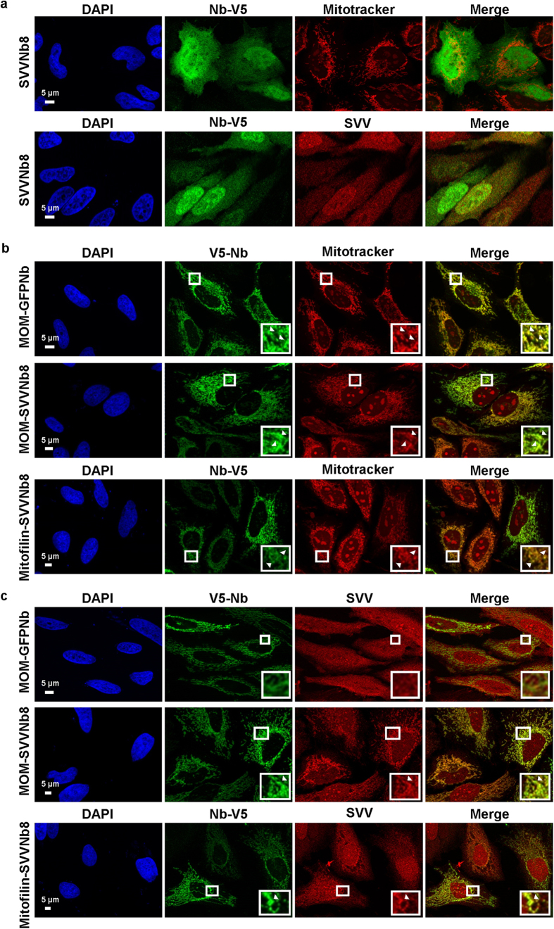
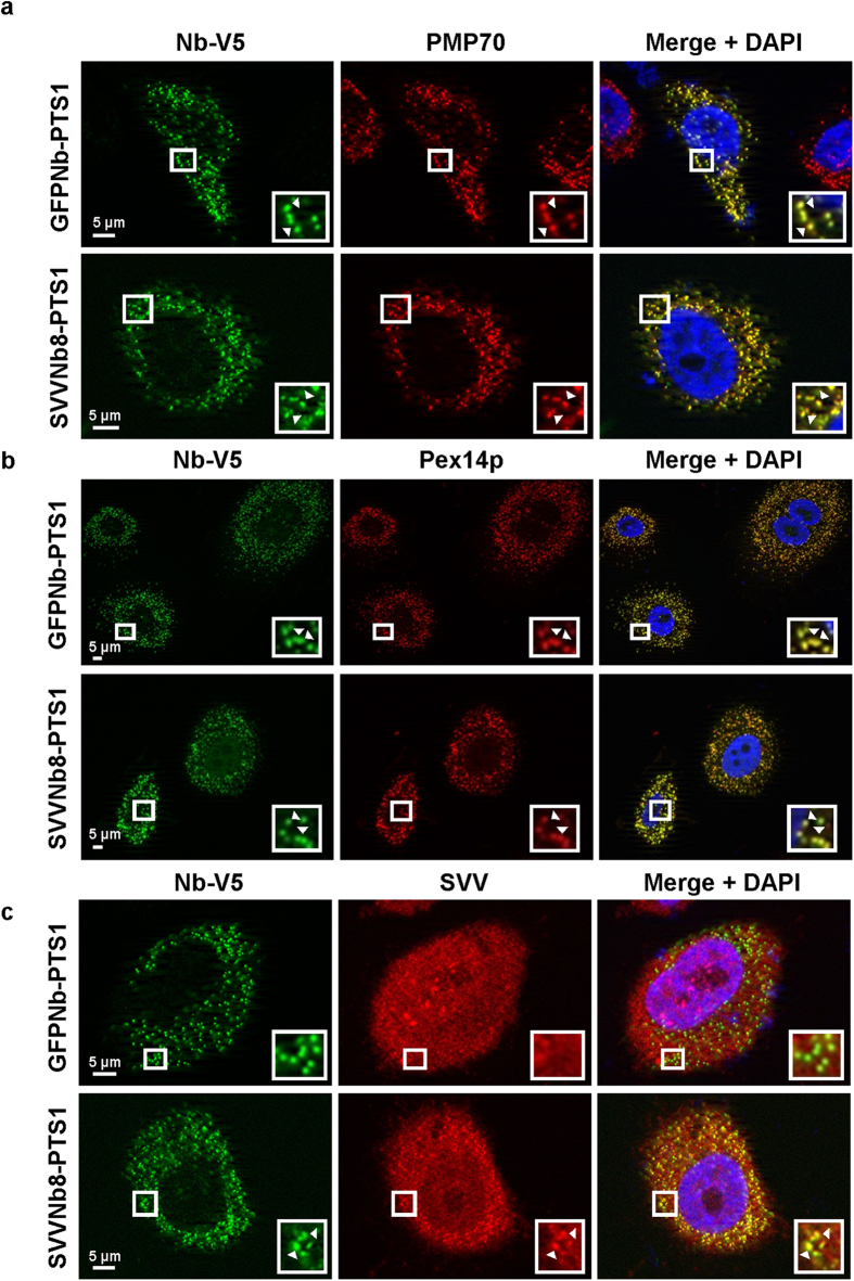
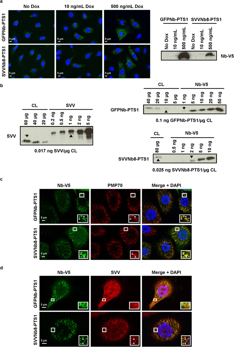
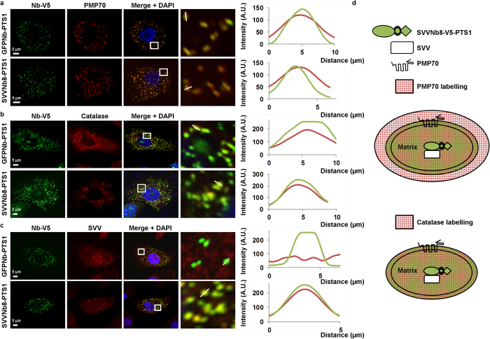
Survivin, the smallest member of the inhibitor of apoptosis protein family, plays a central role during mitosis and exerts a cytoprotective function. Survivin is highly expressed in most cancer types and contributes to multiple facets of carcinogenesis. The molecular mechanisms underlying its highly diverse functions need to be extensively explored, which is crucial for rational design of future personalized therapeutics. In this study, we have generated an alpaca survivin nanobody (SVVNb8) that binds with low nanomolar affinity to its target. When expressed as an intrabody in HeLa cells, SVVNb8 faithfully tracks survivin during different phases of mitosis without interfering with survivin function. Furthermore, coupling SVVNb8 with a subcellular delocalization tag efficiently redirects endogenous survivin towards the nucleus, the cytoplasm, peroxisomes and even to the intermembrane space of mitochondria where it presumably interacts with resident mitochondrial survivin. Based on our findings, we believe that SVVNb8 is an excellent instrument to further elucidate survivin biology and topography, and can serve as a model system to investigate mitochondrial and peroxisomal (survivin) protein import.


Figures

Figure 1
Figure 1. Validation and characterization of SVV nanobodies.
(a) Phylogenetic tree of 28 SVV nanobodies representing their interrelations. The nanobodies can be classified into 22 different groups according to their amino acid sequence, of which 3 groups (depicted by horizontal solid lines) contain several members. Δ represents non-functional recombinant nanobodies. Unlabelled nanobodies bind SVV recombinantly. Nanobodies were additionally classified according to their intracellular binding capacity. This results in the following candidates for further use: Nb8-11, Nb14-15, Nb19, Nb22 and Nb27. (b) Pull-down experiment of endogenous SVV from HeLa cervix cancer cell lysate with recombinant HA-tagged nanobodies by means of anti-HA agarose beads. A negative control was included using HeLa cell lysate with anti-HA agarose beads only (C1). Cells expressing CapGNb2, a nanobody targeting the capping protein CapG, were used as a negative control (C2). Nanobodies were blotted with anti-HA antibody. CL = crude lysate. Full-length blots are presented in Supplementary Figure S4. (c) Immunoprecipitation experiment of endogenous SVV in HEK293T cells transfected with EGFP-tagged nanobodies, by means of an anti-GFP antibody. Two negative controls were included using cell lysates with EGFP only (C1) or CapGNb4-EGFP expression (C2). An anti-GFP antibody was used to visualize the nanobodies. LC = light chain, HC = heavy chain of the anti-GFP antibody. CL = crude lysate. *Depicts breakdown products of the EGFP-tagged nanobody construct. Full-length blots are presented in Supplementary Figure S4.
Figure 2
Figure 2. Detailed binding characterization of SVVNb8.
(a) Epitope mapping experiment with GST only (control), GST-tagged full-length SVV (1), the BIR domain without (2) or with (3) dimer interfaces or the α-helix domain (4) as depicted at the left. The right upper panel shows the different recombinant GST fusion proteins. A Coomassie-stained gel of the GST-pull-down experiment is shown in the right lower panel. SVVNb8 (*) can only be found together with full-length SVV. (b) Representative blot on lysates of HEK293T, HeLa cervix cancer or PC-3 prostate cancer cells using recombinant His6/STREP-tagged SVVNb8 as Western blot reagent followed by an anti-His6 antibody. A commercial polyclonal anti-SVV antibody (ab469) and a His6/STREP-tagged GFP-targeting nanobody were used as positive and negative control, respectively. SVVNb8 does not detect SVV on Western blot. Full-length blots are presented in Supplementary Figure S4. (c) ITC profile of recombinant His6/STREP-tagged SVVNb8 (injected) with recombinant GST-SVV (sample cell). The upper panel shows raw data of heat release as a function of time, while the lower panel shows the fitted binding curve of total heat release per injection as a function of the molar ratio. SVVNb8 binds SVV with an affinity of Kd = 0.95 ± 0.53 nM and a molar ratio of 2:1 SVVNb8:SVV. (d) Western blot after native PAGE of recombinant purified His6-SVV, GST-SVV and GST-cortactin. GST-cortactin was used as a control and native proteins were detected with anti-SVV (left and middle) or anti-cortactin (right) antibody. Both His6-SVV and GST-SVV can form dimers in solution. *Depicts monomeric protein, **Depicts dimeric protein.
Figure 3
Figure 3. SVVNb8 colocalizes with SVV in different cell cycle stages without affecting cell viability.
(a) Endogenous SVV is typically enriched at the spindle midzone and the midbody between anaphase and cytokinesis, as visualized in HeLa cervix cancer cells (arrowheads Δ). (b) HeLa cervix cancer cells transfected with a GFP-targeting nanobody (control) show no enrichment at the spindle midzone (first panel) or midbody (second panel). On the other hand, SVVNb8 is enriched at the spindle midzone (third panel) or midbody (lower panel) upon expression (boxed areas, arrowheads Δ). Boxed areas are enlarged in the insets. SVVNb8 is also enriched between sister chromatids and at the cell poles (arrows ) during late anaphase. Nuclei were visualized by means of DAPI and nanobodies with anti-V5 antibody. (c) Representative co-immunoprecipitation experiment of endogenous Hsp90AA1-SVV complex in MDA-MB-231 breast cancer cells by means of anti-SVV antibody (D-8) coupled on protein G sepharose beads. A negative control was included using cell lysate with protein sepharose beads only (lane 3), representing the level of aspecific SVV binding on naked beads. Crude lysates were incubated with GFPNb (lane 1) or SVVNb8 (lane 2) prior to antibody-mediated SVV pull-down, or no nanobody was added (lane 4, positive control). Hsp90AA1 only co-precipitates when beads are specifically enriched for SVV (lane 4). SVVNb8 does not affect the interaction between Hsp90AA1 and SVV (lane 2), nor does GFPNb (lane 1). Only SVVNb8 co-precipitates with the complex (lane 2). Lane 5 and lane 6 contain 1 μg of GFPNb and SVVNb8, respectively. An anti-His6 antibody was used to visualize the nanobodies. Longer exposure time was needed to detect SVV and Hsp90AA1 in crude lysate (CL) than in other conditions. Full-length blots are presented in Supplementary Figure S4. (d) XTT cell viability assay on HeLa cells transiently expressing EGFP (control) or SVVNb8-EGFP. Graphs represent mean net absorbance of formazan dye (dotted line) with range (three times three independent measurements; n = 3). P0.05-value was determined by a Mann-Whitney Rank Sum Test. SVVNb8-EGFP expression does not significantly alter cell viability compared to EGFP expression (p = 0.40).
Figure 4
Figure 4. SVVNb8 redirects endogenous SVV in and out of the nucleus.
Representative epifluorescence images of HeLa cervix cancer cells transiently expressing EGFP-tagged SVVNb8 without (a) or with an nuclear export signal (b) or nuclear localization signal (c). CapGNb4 was used as a negative control. The nanobody and endogenous SVV are distributed over the whole cell body (a). The nuclear export signal (NES) excludes both nanobodies from the nucleus, with only SVVNb8-NES resulting in delocalization of SVV (b). The nuclear localization signal (NLS) guides both nanobodies towards the nucleus, but only SVVNb8-NLS results in nuclear import of SVV (c). Nuclei are visualized with DAPI.
Figure 5
Figure 5. SVVNb8 captures endogenous SVV at mitochondria.
Representative epifluorescence images of HeLa cervix cancer cells transiently expressing SVVNb8 (a) equipped with a MOM or mitofilin-tag (b,c). MOM-tagged GFP nanobody was used as a negative control. The SVV nanobody and SVV are distributed throughout the cell without enrichment at the mitochondria (a). MOM or mitofilin-tagged control or SVV nanobodies become enriched at the mitochondria, labelled with Mitotracker (b). However, SVV only adopts a mitochondria-like pattern when MOM or mitofilin-tagged SVVNb8 is expressed (c). Nuclei were visualized with DAPI and nanobodies with anti-V5 antibody. Boxed areas are enlarged in the bottom right insets and arrowheads Δ indicate areas of colocalization.
Figure 6
Figure 6. The PTS1-tagged nanobody redirects SVV towards the peroxisomes.
Representative epifluorescence images of PC-3 prostate cancer cells transiently expressing PTS1-tagged GFP nanobody (control) or SVVNb8. Both nanobodies colocalize with the peroxisomal markers PMP70 (a) and Pex14p (b). Only SVVNb8-V5/PTS1, but not the control nanobody, is able to induce SVV delocalization to a peroxisome-like pattern (c). Nuclei are visualized with DAPI and nanobodies with anti-V5 antibody. Boxed areas are enlarged in the bottom right insets and arrowheads Δ indicate areas of colocalization.
Figure 7
Figure 7. Characterization of stable PC-3 cell lines with Dox-inducible expression of PTS1-tagged GFP nanobody or SVVNb8.
(a) Representative epifluorescence images of stable PC-3 cells upon addition of 0 ng/mL, 10 ng/mL or 500 ng/mL Dox for 24 h showing inducibility and concentration-dependence of PTS1-tagged nanobody expression (left). Nuclei are visualized with DAPI and nanobodies with anti-V5 antibody. At the right, a Western blot is depicted, confirming inducibility and concentration-dependence of PTS1-tagged nanobody expression. Full-length blot is presented in Supplementary Figure S4. (b) Determination of the amount of nanobody and endogenous SVV in the generated stable cell lines induced with 500 ng/mL Dox by comparing Western blot analysis on crude lysate (CL) with recombinant standards (His6-SVV and LPLNb5-V5, a nanobody targeting the actin-bundling protein L-plastin). Nanobodies were blotted with anti-V5 antibody. Arrowheads Δ indicate the estimated amount of protein corresponding to the level in the lysate, resulting in the ratios depicted at the bottom. Approximately six times more PTS1-tagged GFP nanobody is present compared to endogenous SVV. Conversely, PTS1-tagged SVVNb8 is expressed at an approximately 1:1 molar ratio to endogenous SVV. Full-length blots are presented in Supplementary Figure S4. (c) Representative epifluorescence images of PC-3 prostate cancer cells stably expressing PTS1-tagged GFP nanobody (control) or SVVNb8 upon induction with 500 ng/mL Dox. Both the control nanobody and SVVNb8-V5/PTS1 colocalize with the peroxisomal marker PMP70. (d) Only SVVNb8-V5/PTS1 is able to delocalize SVV towards the peroxisomes. Nuclei are visualized with DAPI and nanobodies with anti-V5 antibody. Boxed areas are enlarged in the bottom right insets and arrowheads Δ indicate regions of colocalization.
Figure 8
Figure 8. SVVNb8 localizes at the peroxisomal matrix and redistributes SVV accordingly.
Representative confocal images of PC-3 prostate cancer cells expressing PTS1-tagged GFP nanobody (control) or SVVNb8 upon 24 h induction with 500 ng/mL Dox. Both the control and SVVNb8 colocalize with the peroxisomal marker PMP70 (a), although PMP70 is additionally enriched at a region surrounding the nanobody. Colocalization studies with catalase on the other hand, reveal intensity overlap with the nanobody (b). Colocalization of SVV with the GFP nanobody does not occur, but is complete with the SVVNb8 (c). Nuclei are visualized with DAPI and nanobodies with anti-V5 antibody. Boxed areas are enlarged at the right. Graphs represent corresponding intensity profiles generated through the white solid lines. (d) Schematic representation of the different components at the peroxisomes. PMP70 is a transmembrane protein and its signal colocalizes with and additionally surrounds the nanobody signal. Catalase on the other hand is an internal peroxisomal component and colocalizes completely with SVVNb8. SVVNb8 signal colocalizes with the SVV signal.

References

    1. Ambrosini G., Adida C. & Altieri D. C. A novel anti-apoptosis gene, survivin, expressed in cancer and lymphoma. Nat. Med. 3, 917–921, 10.1038/nm0897-917 (1997). - DOI - PubMed
    1. Verdecia M. A. et al.. Structure of the human anti-apoptotic protein survivin reveals a dimeric arrangement. Nat. Struct. Biol. 7, 602–608 (2000). - PubMed
    1. Pavlyukov M. S. et al.. Survivin Monomer Plays an Essential Role in Apoptosis Regulation. J. Biol. Chem. 286, 23296–23307, 10.1074/jbc.M111.237586 (2011). - DOI - PMC - PubMed
    1. Li F. Z. et al.. Control of apoptosis and mitotic spindle checkpoint by survivin. Nature 396, 580–584 (1998). - PubMed
    1. Carvalho A., Carmena M., Sambade C., Earnshaw W. C. & Wheatley S. P. Survivin is required for stable checkpoint activation in taxol-treated HeLa cells. J. Cell Sci. 116, 2987–2998, 10.1242/jcs.00612 (2003). - DOI - PubMed

Publication types

MeSH terms