

STED and STORM Superresolution Imaging of Primary Cilia
- PMID: 27514922
- PMCID: PMC6865270
- DOI: 10.1007/978-1-4939-3789-9_11
STED and STORM Superresolution Imaging of Primary Cilia
Abstract
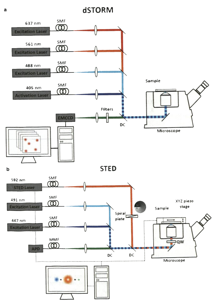
The characteristic lengths of molecular arrangement in primary cilia are below the diffraction limit of light, challenging structural and functional studies of ciliary proteins. Superresolution microscopy can reach up to a 20 nm resolution, significantly improving the ability to map molecules in primary cilia. Here we describe detailed experimental procedure of STED microscopy imaging and dSTORM imaging, two of the most powerful superresolution imaging techniques. Specifically, we emphasize the use of these two methods on imaging proteins in primary cilia.
Keywords: Ciliary protein; Diffraction limit; Fluorophore; Primary cilium; STED; STORM; Superresolution microscopy.
Figures
Similar articles
-
Superresolution STED microscopy reveals differential localization in primary cilia.Cytoskeleton (Hoboken). 2013 Jan;70(1):54-65. doi: 10.1002/cm.21090. Epub 2012 Nov 16. Cytoskeleton (Hoboken). 2013. PMID: 23125024
-
Practical method for superresolution imaging of primary cilia and centrioles by expansion microscopy using an amplibody for fluorescence signal amplification.Mol Biol Cell. 2020 Sep 15;31(20):2195-2206. doi: 10.1091/mbc.E20-04-0250. Epub 2020 Jul 29. Mol Biol Cell. 2020. PMID: 32726175 Free PMC article.
-
CLEM Methods for Studying Primary Cilia.Methods Mol Biol. 2016;1454:193-202. doi: 10.1007/978-1-4939-3789-9_12. Methods Mol Biol. 2016. PMID: 27514923
-
Nanoscale imaging by superresolution fluorescence microscopy and its emerging applications in biomedical research.Crit Rev Biomed Eng. 2013;41(4-5):281-308. doi: 10.1615/critrevbiomedeng.2014010448. Crit Rev Biomed Eng. 2013. PMID: 24941410 Review.
-
Technical review: types of imaging-direct STORM.Anat Rec (Hoboken). 2014 Dec;297(12):2227-31. doi: 10.1002/ar.22960. Epub 2014 Jul 4. Anat Rec (Hoboken). 2014. PMID: 24995970 Review.
Cited by
-
Retinal primary cilia and their dysfunction in retinal neurodegenerative diseases: beyond ciliopathies.Mol Med. 2024 Jul 26;30(1):109. doi: 10.1186/s10020-024-00875-y. Mol Med. 2024. PMID: 39060957 Free PMC article. Review.
-
Primary cilia biogenesis and associated retinal ciliopathies.Semin Cell Dev Biol. 2021 Feb;110:70-88. doi: 10.1016/j.semcdb.2020.07.013. Epub 2020 Jul 31. Semin Cell Dev Biol. 2021. PMID: 32747192 Free PMC article. Review.
-
Two separate functions of NME3 critical for cell survival underlie a neurodegenerative disorder.Proc Natl Acad Sci U S A. 2019 Jan 8;116(2):566-574. doi: 10.1073/pnas.1818629116. Epub 2018 Dec 26. Proc Natl Acad Sci U S A. 2019. PMID: 30587587 Free PMC article.
References
-
- Chih B, Liu P, Chinn Y, Chalouni C, Komuves LG et al. (2011) A ciliopathy complex at the transition zone protects the cilia as a privileged membrane domain. Nat Cell Biol 14:61–72 - PubMed
Publication types
MeSH terms
Grants and funding
LinkOut - more resources
Full Text Sources
Other Literature Sources
