Review
. 2016 Aug 15;473(16):2439-52.
doi: 10.1042/BCJ20160005.

Mechanisms of Hsp90 regulation

Affiliations
Review

Mechanisms of Hsp90 regulation

Chrisostomos Prodromou. Biochem J. .

Abstract

Heat shock protein 90 (Hsp90) is a molecular chaperone that is involved in the activation of disparate client proteins. This implicates Hsp90 in diverse biological processes that require a variety of co-ordinated regulatory mechanisms to control its activity. Perhaps the most important regulator is heat shock factor 1 (HSF1), which is primarily responsible for upregulating Hsp90 by binding heat shock elements (HSEs) within Hsp90 promoters. HSF1 is itself subject to a variety of regulatory processes and can directly respond to stress. HSF1 also interacts with a variety of transcriptional factors that help integrate biological signals, which in turn regulate Hsp90 appropriately. Because of the diverse clientele of Hsp90 a whole variety of co-chaperones also regulate its activity and some are directly responsible for delivery of client protein. Consequently, co-chaperones themselves, like Hsp90, are also subject to regulatory mechanisms such as post translational modification. This review, looks at the many different levels by which Hsp90 activity is ultimately regulated.

Keywords: HSF1; Hsp90; chaperones; co-chaperones; heat-shock response; post-translational modification.


Figures

Figure 1
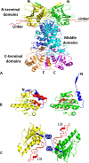
Figure 1. Structure and conformational change in Hsp90
(A) The Hsp90 dimer in a closed conformation involving transient dimerization of the N-terminal domains. N-terminal domains, yellow and green; middle domains, blue and cyan; C-terminal domains, orange and magenta; charged linker, red. (B) Conformation of the lid and N-terminal segment of the N-terminal domains of Hsp90 in the open undimerized state (left-hand panel, yellow) and the closed dimerized state (right-hand panel, green). Lids, red; N-terminal segment, blue. (C) The closed transient N-terminal dimerized state of Hsp90 (yellow and green). Lids, red; N-terminal segment, blue. N-terminal dimerization involves movement of the N-terminal segments of the N-terminal domains and association with the closed lid segments and with the neighbouring N-terminal domains.
Figure 2
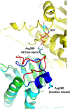
Figure 2. Conformation of the catalytic loop of Hsp90
The N-terminal domain of Hsp90 is shown in yellow. The middle domain is represented by two superimposed molecules of Hsp90 (cyan and green), one with a closed inactive catalytic loop (blue) and the other with an open active state (red) that interacts with the bound ATP, which is shown as a stick model. Arg380 of the catalytic loop is either interacting with the ATP (active state) or is held in an inactive state. Broken blue lines represent hydrogen bonds.
Figure 3
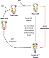
Figure 3. The Hsp90 chaperone cycle
ATP binding triggers transient N-terminal dimerization, through conformational changes in the N-terminal domain of Hsp90 including those of the lid and N-terminal segment, and association with the catalytic loop of the middle domain. These motions act co-operatively to form the catalytically active closed state of Hsp90. Aha1, can accelerate the formation of this closed state by modulating the catalytic loop to an active open state. Once ATP is hydrolysed the N-terminal domains separate, the open inactive state of Hsp90 is formed and ADP is released. Hsp90 is now ready to enter the next cycle.
Figure 4
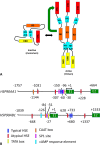
Figure 4. Domain structure of mammalian HSF1 and the promoter and upstream control elements of Hsp90-encoding genes
(A) The domain structure of HSF1. The N-terminal DBD consists of the first 110 amino acids and is followed by the HR-A/B trimerization domain consisting of amino acids 130–203. The RD is encompassed by residues 221–383 and is followed by the HR-C region, residues 384–409, which negatively regulate the HR-A/B trimerization domain. The CTA domain consists of residues 410–529 and is negatively regulated by the RD (green arrow). Activation of HSF1 involves trimerization through the HR-A/B domains. Yeast HSF1 differs in that it also possesses an N-terminal transactivation domain that negatively regulates the RD. (B) Promoter and regulatory regions of human Hsp90-encoding genes. Sequences start from 5′ upstream regions, −1757 for HSP90AA1 and −1039 for HSP90AB1, and end at the second exon. The approximate locations of various control elements are indicated and can be identified from the key. E1 and E2 are exons 1 and 2. The UPE (−125 to −37 bp) region of HSP90AA1 confers a 10-fold up-regulation of the core promoter. The core promoter (−36 to +37 bp) of HSP90AB1 confers constitutive expression, and the two typical HSE are responsible for maintaining a high level of constitutive expression.
Figure 5
Figure 5. Integration of signalling pathways in the control of Hsp90 expression
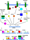
(A) Known signalling pathways that affect Hsp90 expression. The co-activator Daxx is not shown in the scheme, but is known to promote the activation of HSF1 [69]. (B) Possible binding scenario for transcriptional activators and cofactors that regulate Hsp90 transcription. IFN-γ, interferon-γ; IL-R, interleukin receptor; JAK, Janus kinase; MAPK, mitogen-activated protein kinase.
Figure 6
Figure 6. Post-translational modification of Hsp90
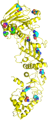
A single monomer of yeast Hsp90 (yellow) is shown in cartoon format. Amino acid residues from yeast and human Hsp90 that are post-translationally modified are shown as spheres and mapped to the correct location on yeast Hsp90. Modified amino acid residues for Hsp90α Ser5, Ser7 and Ser234 and Thr725 and Hsp90β Ser255 that are not represented in the yeast structure are omitted. Green spheres, amino acids that are phosphorylated; cyan spheres, amino acids that are acetylated, magenta spheres, amino acids that are SUMOylated; gold spheres, amino acids that are nitrosylated.
Figure 7
Figure 7. Structure of Hsp90–Aha1 and Hsp90–Cdc37p50 co-chaperone complexes
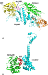
(A) Structure of the Hsp90–Aha1 complex by superimposition of the middle domain of Hsp90 (cyan) in complex with Aha1 (green) on to the full-length structure of Hsp90 (N-terminus, yellow; C-terminus, gold). The binding of Aha1 causes the catalytic loop of Hsp90 (magenta) to move to its open state and allows Arg380 to interact with the γ-phosphate of ATP (green stick representation). Broken blue lines represent hydrogen bonds. (B) Structure of the N-terminal domain of Hsp90 (green) in complex with the C-terminal domain of Cdc37p50 (cyan). Cdc37p50 binds to the lid segment (red) of the N-terminal domains of Hsp90, preventing them from conformational movements that are required for the formation of the catalytically active state through N-terminal dimerization.
Figure 8
Figure 8. Co-chaperone pathways that modulate Hsp90 ATPase activity
Cdc37p50 binds to the lids and prevents molecular rearrangement of Hsp90. HOP appears to interact with N-terminal segment of Hsp90 and thus may prevent N-terminal dimerization. Aha1 is able to interact with possibly all of the structural elements that lead to co-operative N-terminal dimerization of Hsp90. Sba1 interacts with the lid and N-terminal domains of Hsp90 and stabilizes Hsp90 in a closed state that displays a lower rate of ATP hydrolysis. Sgt1, together with Rar1, is unusual in that it activates Hsp90 in an open state and leads to a stable ADP-bound complex. Red and blue arrows indicate a mechanism resulting in the inhibition and activation of ATPase activity respectively. Broken blue arrows indicate interactions that might occur. The cyan arrow indicates a means by which the rate of ATPase activity is decreased. The green arrows indicate the co-operative nature of N-terminal dimerization.
Figure 9
Figure 9. Structure of Hsp90–Rar1–Sgt1 and Hsp90–Rar1–Sba1 co-chaperone complexes
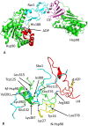
(A) Structure of the N-terminal domains of Hsp90 (green) in complex with the CS domain of Sgt1 (magenta) and the CHORD II domain of Rar1 (cyan). Recruitment of Rar1 into the Hsp90 complex stimulates ATP hydrolysis producing a stable ADP-bound Hsp90 complex. The lid segment is shown in red and bound ADP in blue stick representation. Broken blue lines represent hydrogen bonds. (B) Structure of the closed conformation of Hsp90 in complex with Sba1 (cyan). Hsp90 is represented by the lid (red), the N-terminal segment of the N-terminus (yellow) and a segment of the middle domain (green). The bound ATP is shown as a yellow stick representation. Arg380 is seen to interact with the γ-phosphate of ATP in the catalytically active state of Hsp90. Broken blue lines represent hydrogen bonds.

References

    1. Picard D. Heat-shock protein 90, a chaperone for folding and regulation. Cell Mol Life Sci. 2002;59:1640–1648. doi: 10.1007/PL00012491. - DOI - PMC - PubMed
    1. Whitesell L., Lindquist S.L. HSP90 and the chaperoning of cancer. Nat Rev Cancer. 2005;5:761–772. doi: 10.1038/nrc1716. - DOI - PubMed
    1. Taipale M., Jarosz D.F., Lindquist S. HSP90 at the hub of protein homeostasis: emerging mechanistic insights. Nature reviews. Molecular cell biology. 2010;11:515–528. doi: 10.1038/nrm2918. - DOI - PubMed
    1. Finka A., Goloubinoff P. Proteomic data from human cell cultures refine mechanisms of chaperone-mediated protein homeostasis. Cell Stress Chaperones. 2013;18:591–605. doi: 10.1007/s12192-013-0413-3. - DOI - PMC - PubMed
    1. Ammirante M., Rosati A., Gentilella A., Festa M., Petrella A., Marzullo L., Pascale M., Belisario M.A., Leone A., Turco M.C. The activity of hsp90 alpha promoter is regulated by NF-kappa B transcription factors. Oncogene. 2008;27:1175–1178. doi: 10.1038/sj.onc.1210716. - DOI - PubMed

Publication types

MeSH terms

Substances

LinkOut - more resources