. 2016 Aug 12:6:31545.
doi: 10.1038/srep31545.

Elimination of microglia improves cognitive function following cranial irradiation

Affiliations

Elimination of microglia improves cognitive function following cranial irradiation

Munjal M Acharya et al. Sci Rep. .

Abstract

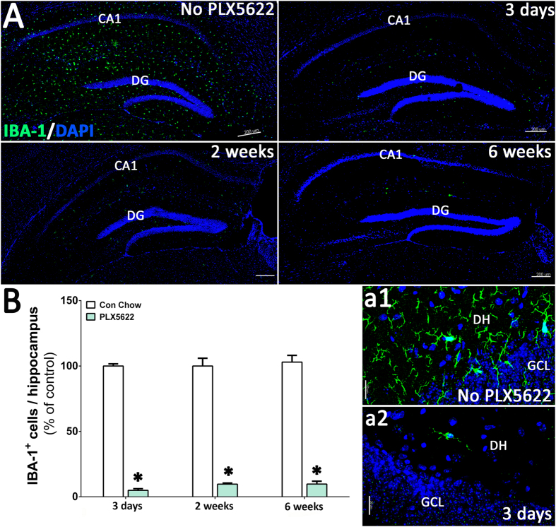
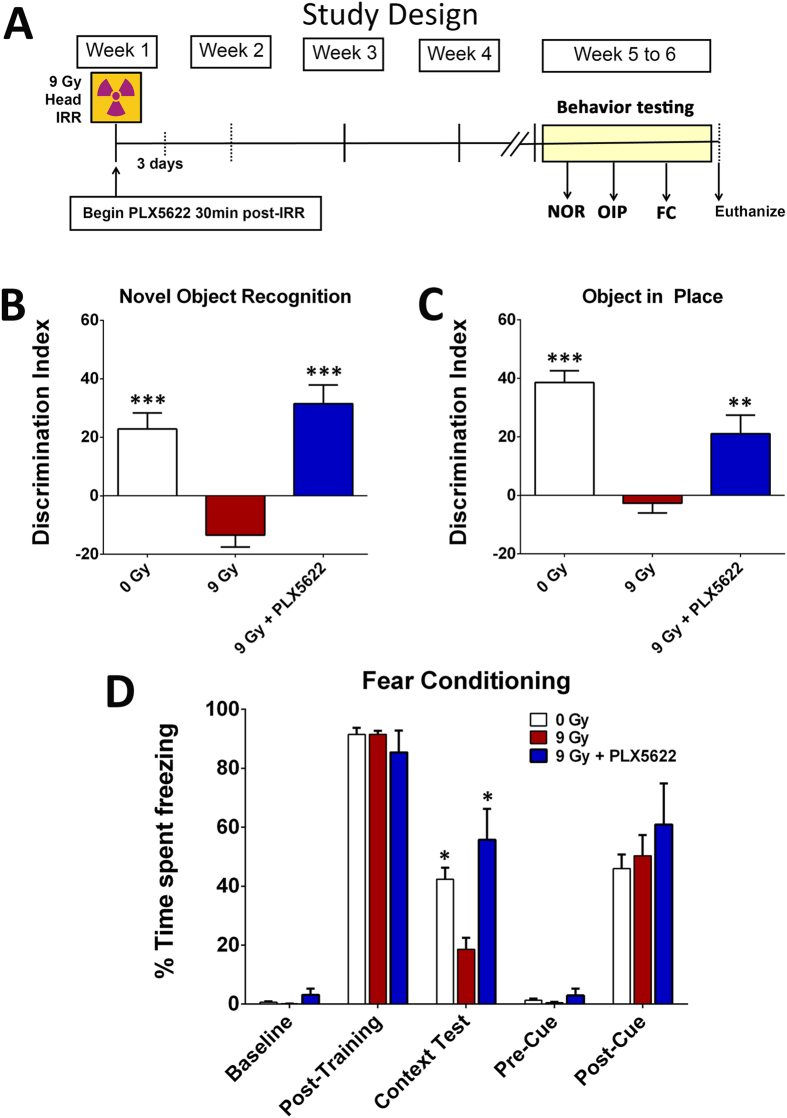
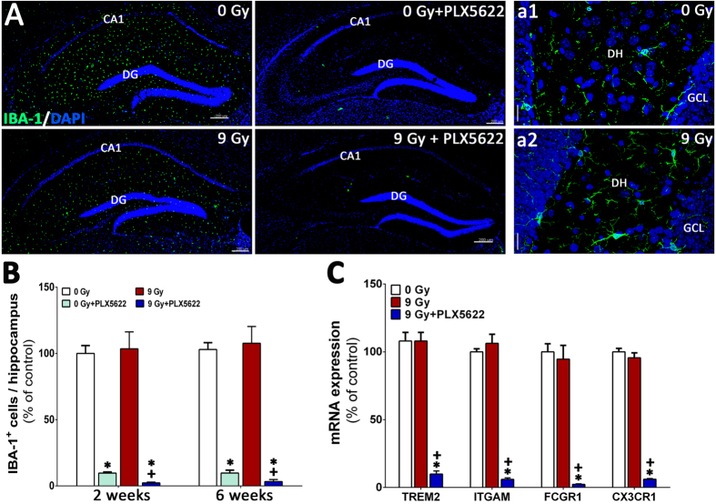
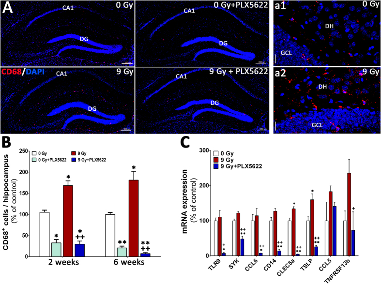
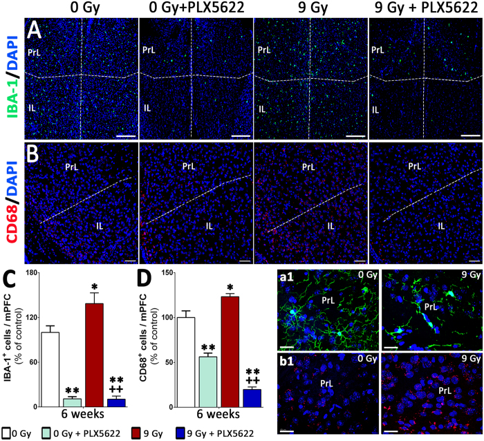
Cranial irradiation for the treatment of brain cancer elicits progressive and severe cognitive dysfunction that is associated with significant neuropathology. Radiation injury in the CNS has been linked to persistent microglial activation, and we find upregulation of pro-inflammatory genes even 6 weeks after irradiation. We hypothesize that depletion of microglia in the irradiated brain would have a neuroprotective effect. Adult mice received acute head only irradiation (9 Gy) and were administered a dietary inhibitor (PLX5622) of colony stimulating factor-1 receptor (CSF1R) to deplete microglia post-irradiation. Cohorts of mice maintained on a normal and PLX5662 diet were analyzed for cognitive changes using a battery of behavioral tasks 4-6 weeks later. PLX5622 treatment caused a rapid and near complete elimination of microglia in the brain within 3 days of treatment. Irradiation of animals given a normal diet caused characteristic behavioral deficits designed to test medial pre-frontal cortex (mPFC) and hippocampal learning and memory and caused increased microglial activation. Animals receiving the PLX5622 diet exhibited no radiation-induced cognitive deficits, and exhibited near complete loss of IBA-1 and CD68 positive microglia in the mPFC and hippocampus. Our data demonstrate that elimination of microglia through CSF1R inhibition can ameliorate radiation-induced cognitive deficits in mice.


Conflict of interest statement

B.L.W. is an employee of Plexxikon Inc. The remaining authors declare no competing financial interests.

Figures

Figure 1
Figure 1. CSF1 receptor (CSF1R) inhibition eliminates microglia from the adult mouse brain.
Six month old, male wild type mice were administered control or PLX5622 rodent chow for 3 days, 2 weeks or 4–6 weeks. (A) Immunofluorescence staining and laser scanning confocal microcopy was performed on representative brain sections from each group for the microglial marker IBA-1. PLX5622 treatment led to rapid elimination of IBA-1+ microglia throughout the brain, with nearly complete elimination after 6 weeks of treatment. (a1,a2) Representative high-resolution (60×) confocal micrographs from the hippocampal dentate hilus (DH) and granule cell layer (GCL) are shown for the control and PLX5622 treated groups for 3 day treatment. (B) 3D algorithm-based z stack deconvolution (AutoQuant) and quantification (Imaris) of the number of IBA-1+ microglial cell bodies from the hippocampal region show robust decreases in microglial numbers for all the treatment time points. Data are presented as mean ± SEM (N = 4 animals/group). P values are derived from ANOVA and Bonferroni’s multiple comparisons test. *P < 0.001 compared with control group. Scale bars: 200 μm (A) and 20 μm (a1,a2).
Figure 2
Figure 2. Treatment with CSF1R inhibitor did not affect cognitive function in the intact animals.
Unirradiated (0 Gy) adult mice received PLX5622 (1200 mg/kg) or control chow for one month and tested on the hippocampal- and medial pre-frontal cortex (mPFC)-dependent cognitive function tasks (novel object recognition, NOR; object in place, OIP and fear conditioning, FC, A–C). The discrimination Index (DI), calculated as ([Novel location exploration time/Total exploration time] − [Familiar location exploration time/Total exploration time]) × 100. PLX5622 treatment did not affect cognitive function as indicated by indistinguishable performance of 0 Gy + PLX5622 and 0 Gy groups. Data are presented as mean ± SEM (N = 8–10 mice/group).
Figure 3
Figure 3. CSF1R inhibition prevents cranial radiation-induced cognitive dysfunction.
(A) Schematic of study design: 6 month old wild type male mice were irradiated (0 or 9 Gy, head only) and administered CSF1R inhibitor PLX5622 in rodent chow after irradiation (IRR) and continued on diet till the end of study. Animals that received control chow served as vehicle group. A small cohort of mice was euthanized at 3 days and 2 weeks of PLX5622 treatments for assessment of microglial elimination. One month post-IRR and PLX5622 treatment (week 5 to 6), mice were tested on spatial and episodic memory retention using the novel object recognition (NOR) and object in place (OIP) tasks followed by fear conditioning (FC) task. (BD) The tendency to explore novel location(s) was derived from the Discrimination Index (DI), calculated as ([Novel location exploration time/Total exploration time] − [Familiar location exploration time/Total exploration time]) × 100. Cranial irradiation (9 Gy) show significant behavioral deficits on the NOR and OIP tasks compared to controls (0 Gy) as indicated by impaired preference to novel object (B) or place (C). Irradiated animals receiving CSF1R inhibitor (9 Gy + PLX5622) show significant preference for the novelty when compared with irradiated (9 Gy) animals receiving vehicle. (D) CSF1R inhibitor improves behavior on the hippocampal-dependent contextual fear-conditioning task. The baseline freezing levels were comparable among groups, and all groups showed elevated freezing behavior following a series of 5 tone-shock pairings (post-training bars). The context test was administered 24 hour later, and irradiated mice (9 Gy) showed significantly decreased freezing compared to controls (0 Gy). Irradiated animals receiving PLX5622 diet (9 Gy + PLX5622) showed a significant elevation in freezing behavior that was indistinguishable from the 0 Gy group. After the initial training phase (48 hours), the context was changed that resulted in a considerable reduction in freezing behavior (Pre-Cue bars) that was restored following the tone sound (Post-Cue test bars), indicating intact amygdala function in all groups. Data are presented as mean ± SEM (N = 8–10 mice/group). P values are derived from ANOVA and Bonferroni’s multiple comparisons test. ***P < 0.001; **P < 0.01, *P < 0.05 compared with the 9 Gy group.
Figure 4
Figure 4. CSF1R inhibition eliminates IBA-1+ microglia from the unirradiated and irradiated hippocampus.
(A) Immunofluorescence staining and laser scanning confocal microscopy demonstrates that PLX5622 treatment for 6 weeks eliminates ~90% of IBA-1+ microglia from the control (0 Gy + PLX5622) and irradiated (9 Gy + PLX5622) brains. (a1,a2) Representative high-resolution (60×) confocal micrographs from the hippocampal dentate hilus (DH) and granule cell layer (GCL) are shown for the 0 and 9 Gy mice that received control chow. (B) 3D algorithm-based quantification (Autoquant and Imaris) of IBA-1+ microglia show almost complete elimination (90–96%) of microglia in the control and irradiated brains of animals treated with PLX5622 (0 Gy + PLX5622 and 9 Gy + PLX5622) at 2 week and 6 week post-treatments. (C) Gene expression analysis of microglial markers from whole brains derived from a separate cohort of irradiated animals (0 and 9 Gy) received 1 week of PLX5622 treatment at 4 week post-irradiation show 80–90% reduction in mRNA levels after treatment with PLX5622. Data are presented as mean ± SEM (N = 4 mice/group). P values are derived from ANOVA and Bonferroni’s multiple comparisons test. *P < 0.001 compared with 0 Gy group and +P < 0.001 compared with the 9 Gy group. Scale bars: 200 μm (A) and 20 μm (a1,a2).
Figure 5
Figure 5. CSF1R inhibition reduces CD68+ activated microglia from the irradiated hippocampus.
(A) Immunofluorescence staining show that PLX5622 treatment for 6 weeks reduces ~70% of CD68+ activated microglia from the control (0 Gy + PLX5622) and irradiated (9 Gy + PLX5622) brains. (a1,a2) Representative high-resolution (60×) z stacks show ramified microglial morphology in the irradiated hippocampal dentate hilus (DH) and granule cell layer (GCL) compared to 0 Gy mice that received control chow. (B) Quantification (Autoquant and Imaris) of CD68+ activated microglia indicated an 80–90% reduction in the control and irradiated brains receiving PLX5622 (0 Gy + PLX5622 and 9 Gy + PLX5622) at 2 week and 6 week time points. (C) Analysis of pro-inflammatory markers from whole brains derived from irradiated mice (0 and 9 Gy) treated with PLX5622 for 1 week at 4 week post-irradiation show radiation-induced elevation in gene expression that was reduced significantly by PLX5622 treatment. Data are presented as mean ± SEM (N = 4 mice/group). P values are derived from ANOVA and Bonferroni’s multiple comparisons test. *P < 0.01; **P < 0.001 compared with 0 Gy group and +P < 0.01; ++P < 0.01compared with 9 Gy group. Scale bars: 200 μm (A) and 20 μm (a1,a2).
Figure 6
Figure 6. Microglial depletion in the medial pre-frontal cortex by CSF1R inhibition.
(A,B) Immunofluorescence staining and laser scanning confocal microscopy for the IBA-1+ and CD68+ cells show that cranial irradiation lead to significant elevation in microglial number (40% and 25% respectively) in the pre-limbic (PrL) and infra-limbic (IL) cortices of the medial pre-frontal cortex (mPFC). (a1,b1) Representative high-resolution (60×) z stacks showed characteristic activated microglial morphology (IBA-1, a1 and CD68, b1) in the irradiated PrL compared to 0 Gy group. (C,D) Treatment with PLX5622 for 6 weeks eliminates 80–90% of IBA-1+ and CD68+ microglia from the control (0 Gy + PLX5622) and irradiated (9 Gy + PLX5622) mPFC. Data are presented as mean ± SEM (N = 4 mice/group). P values are derived from ANOVA and Bonferroni’s multiple comparisons test. *P < 0.05; **P < 0.001 compared with 0 Gy group and +P < 0.05; ++P < 0.01compared with 9 Gy group. Scale bars: 200 μm (A), 100 μm (B) and 50 μm (a1,b1).

References

    1. Elmore M. R. et al.. Colony-stimulating factor 1 receptor signaling is necessary for microglia viability, unmasking a microglia progenitor cell in the adult brain. Neuron 82, 380–397 (2014). - PMC - PubMed
    1. Erblich B., Zhu L., Etgen A. M., Dobrenis K. & Pollard J. W. Absence of colony stimulation factor-1 receptor results in loss of microglia, disrupted brain development and olfactory deficits. PLoS One 6, e26317 (2011). - PMC - PubMed
    1. Ginhoux F. et al.. Fate mapping analysis reveals that adult microglia derive from primitive macrophages. Science 330, 841–845, doi: 10.1126/science.1194637 (2010). - DOI - PMC - PubMed
    1. Elmore M. R., Lee R. J., West B. L. & Green K. N. Characterizing newly repopulated microglia in the adult mouse: impacts on animal behavior, cell morphology, and neuroinflammation. PLoS One 10, e0122912 (2015). - PMC - PubMed
    1. Mok S. et al.. Inhibition of CSF-1 receptor improves the antitumor efficacy of adoptive cell transfer immunotherapy. Cancer Res 74, 153–161 (2014). - PMC - PubMed

Publication types

MeSH terms

Substances