. 2016 Aug 12:6:31022.
doi: 10.1038/srep31022.

Inhibition of DNA Methyltransferases Blocks Mutant Huntingtin-Induced Neurotoxicity

Affiliations

Inhibition of DNA Methyltransferases Blocks Mutant Huntingtin-Induced Neurotoxicity

Yanchun Pan et al. Sci Rep. .

Erratum in

Abstract

Although epigenetic abnormalities have been described in Huntington's disease (HD), the causal epigenetic mechanisms driving neurodegeneration in HD cortex and striatum remain undefined. Using an epigenetic pathway-targeted drug screen, we report that inhibitors of DNA methyltransferases (DNMTs), decitabine and FdCyd, block mutant huntingtin (Htt)-induced toxicity in primary cortical and striatal neurons. In addition, knockdown of DNMT3A or DNMT1 protected neurons against mutant Htt-induced toxicity, together demonstrating a requirement for DNMTs in mutant Htt-triggered neuronal death and suggesting a neurodegenerative mechanism based on DNA methylation-mediated transcriptional repression. Inhibition of DNMTs in HD model primary cortical or striatal neurons restored the expression of several key genes, including Bdnf, an important neurotrophic factor implicated in HD. Accordingly, the Bdnf promoter exhibited aberrant cytosine methylation in mutant Htt-expressing cortical neurons. In vivo, pharmacological inhibition of DNMTs in HD mouse brains restored the mRNA levels of key striatal genes known to be downregulated in HD. Thus, disturbances in DNA methylation play a critical role in mutant Htt-induced neuronal dysfunction and death, raising the possibility that epigenetic strategies targeting abnormal DNA methylation may have therapeutic utility in HD.


Figures

Figure 1
Figure 1. DNMT inhibitors, decitabine and FdCyd, protect neurons from mutant Htt-induced toxicity in culture.
(A) Schematic of epigenetic drug library screen using a primary neuron model. (B) DIV 5 cortical neurons transduced with Htt-expressing lentivirus were treated with decitabine or DMSO (=0 μM), and subjected to MTS assay at DIV 14. Decitabine increased the viability of Htt-72Q-expressing neurons (ANOVA, *P < 0.0001 vs. Htt-25Q (0 μM), #P < 0.0001 vs. Htt-72Q (0 μM), n = 18). (C) Cortical neurons processed as in (B) were fixed at DIV14 and subjected to NF immunofluorescence. Immunofluorescence intensity was quantified. Decitabine blocked Htt-72Q-induced neurite degeneration (ANOVA, *P < 0.0001 vs. Htt-25Q (0 μM), #P < 0.0001 vs. Htt-72Q (0 μM), n = 11–24). (D) Representative NF immunofluorescence images of transduced neurons treated with decitabine (0.2 μM) or vehicle in (C). Bar, 100 μm. (E) (Left) Cortical neurons processed as in (C) were subjected to nuclear staining (Hoechst 33342). Cell death was assessed by nuclear morphology. Decitabine blocked Htt-72Q-induced cell death (ANOVA, *P < 0.0001 vs. Htt-25Q, #P < 0.0001 vs. Htt-72Q (0 μM), n = 16). (Right) Representative nuclear images of transduced neurons. Arrows show examples of condensed or fragmented nuclei, indicating dead cells. Bar, 50 μm. (F,G) Cortical neurons transduced as in (B) were treated with FdCyd and subjected to MTS assay (F) or NF immunofluorescence (G) at DIV 14. FdCyd increased the viability of Htt-72Q-expressing neurons (ANOVA, *P < 0.0001 vs. Htt-72Q (0 μM), n = 11–24) (F). FdCyd protected neurons from Htt-72Q-induced neurite degeneration (ANOVA, *P < 0.0001 vs. Htt-72Q (0 μM); n = 12–24) (G). (H,I) DIV 4 striatal neurons were transduced and treated with the indicated DNMT inhibitor. Seven days later, neurons were fixed and subjected to NF immunofluorescence. Decitabine and FdCyd protected neurons from mutant Htt-induced neurite degeneration (ANOVA, *P < 0.0001 vs. Htt-25Q (0 μM), #P < 0.0001 vs. Htt-72Q (0 μM), n = 13–17 (H); *P = 0.0002 and #P = 0.003 vs. Htt-72Q (0 μM), n = 9–16 (I)). Data are presented as mean + SEM.
Figure 2
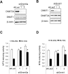
Figure 2. Lentivirus-mediated knockdown of DNMT3A or DNMT1 in primary cortical neurons attenuates mutant Htt-induced toxicity.
(A) DIV 5 cortical neurons were transduced with two Dnmt3a shRNA (1 and 2) or control luciferase (Luci) shRNA lentivirus; 5 days later, cell lysates were subjected to immunoblotting using indicated antibodies. (B) Cortical neurons transduced with two Dnmt1 shRNA (1 and 2) or control LacZ shRNA lentivirus were subjected to immunoblotting as in (A). (C) DIV 5 cortical neurons were co-transduced with Htt-expressing lentivirus along with Dnmt3a or control shRNA lentivirus and were subjected to MTS assay at DIV14. Knockdown of DNMT3A in mutant Htt-expressing neurons was neuroprotective (ANOVA, *P < 0.0001 compared to Htt-72Q plus control RNAi, n = 17–20 wells per group, 5 independent experiments). (D) Cortical neurons co-transduced with Htt lentivirus and Dnmt1 or control shRNA lentivirus were subjected to MTS assay as in (C). Knockdown of DNMT1 in mutant Htt-expressing neurons was neuroprotective (ANOVA, *P < 0.0001 and #P = 0.0001 compared to Htt-72Q plus control RNAi, n = 11–15 wells per group, 4 independent experiments). Data are presented as mean + SEM in (C,D).
Figure 3
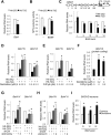
Figure 3. Inhibition of DNMTs restores the expression of Bdnf exon IV and VI transcripts in primary cortical neurons.
(A) DIV 5 cortical neurons were infected with Htt lentivirus. RNA was harvested 5 days later and subjected to qRT-PCR for total Bdnf (coding exon IX) using β-actin and 18S rRNA as reference genes. Htt-72Q decreased the expression of total Bdnf transcripts (Mann-Whitney U test, *P = 0.008 vs. Htt-25Q, n = 5). (B) Cortical neurons transduced as in (A) were cultured with recombinant BDNF (50 ng/ml) and subjected to MTS assay at DIV 14. BDNF increased the viability of Htt-72Q-expressing neurons (ANOVA, *P < 0.0001 vs. Htt-72Q with vehicle, n = 9–15). (C) (Top) Schematic of the mouse Bdnf locus. White boxes, non-coding exons; gray box, coding exon. (Bottom) qRT-PCR was performed as in (A) using exon-specific Bdnf primers. Htt-72Q decreased the expression of exon IV and VI transcripts (Mann-Whitney U test, *P = 0.008 vs. Htt-25Q, n = 5). (DF) Cortical neurons transduced with Htt lentivirus were treated with indicated DNMT inhibitor or vehicle and processed as in (C). gRT-PCR was performed using β-actin and Hprt as reference genes. Both decitabine and FdCyd increased the expression of Bdnf exon IV, VI, and IX transcripts in Htt-72Q-expressing neurons (ANOVA, *P < 0.005 vs. Htt-72Q plus vehicle, n = 5–7 (D); *P < 0.05 vs. Htt-72Q plus vehicle, n = 5 (E); *P < 0.05 vs. Htt-72Q plus vehicle, n = 7 (F)). (G,H) Cortical neurons were co-transduced with lentiviruses expressing Htt and indicated shRNA and processed as in (D). Knockdown of DNMT3A or DNMT1 restored the expression of Bdnf exon IV and VI (ANOVA, *P < 0.05 vs. Htt-72Q plus vehicle, n = 4 (G); *P < 0.01 vs. Htt-72Q plus vehicle, n = 4 (H)). (I) Primary cortical neurons from BACHD mice were treated with decitabine (0.2 μM) or vehicle for 3.5 days. qRT-PCR was performed using β-actin as a reference gene. Decitabine increased expression of Bdnf exon IV, exon VI, and IX transcripts (unpaired t-test, *P < 0.0001 and #P = 0.0012 vs. vehicle treated). Data are presented as mean + SEM.
Figure 4
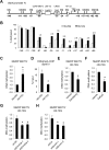
Figure 4. Mutant Htt increases the levels of DNA methylation at Bdnf exon IV regulatory region in primary cortical neurons.
(A) Schematic of the mouse Bdnf exon IV regulatory region near the TSS. The positions of CpG sites are indicated relative to the TSS. (B) DIV 5 primary cortical neurons were infected with lentivirus expressing Htt-25Q or Htt-72Q exon 1 fragment; 5 days later, genomic DNA was purified and subjected to bisulfite sequencing analysis. The data show percentage of cytosine residues that were methylated in Htt25Q- and Htt-72Q-expressing neurons. Increased DNA methylation was found in mutant Htt-expressing neurons compared to WT Htt-expressing neurons. 28–30 clones from 7 independent experiments were analyzed (See Figure S5A for the bisulfite sequencing data from each clone). The number above the black bar (Htt-72Q) represents the fold changes in methylated cytosine relative to the white bar (Htt-25Q) at the indicated position. (C) Genomic DNA was purified from primary cortical neurons transduced as in (B) and subjected to MeDIP with anti-5-mC antibody followed by qPCR. The levels of 5-mC in the exon IV promoter region was higher in Htt-72Q-expressing neurons compared to that in Htt-25Q neurons (Mann-Whitney U test, *P < 0.05, n = 6). (D) Cortical neurons were transduced as in (B) and 5 days later were subjected to ChIP with anti-H3K4me3 antibody. H3K4me3 levels in the exon IV promoter region were lower in Htt-72Q-expressing neurons compared to Htt-25Q neurons. (unpaired t-test, *P < 0.05, n = 5). (E,F) Cortical neurons were processed and subjected to MeDIP as in (C) using Htt-72Q-expressing neurons treated with DNMT inhibitors (0.2 μM) or DMSO. Treatment with decitabine or FdCyd decreased levels of 5-mC in Bdnf promoter IV region (Mann-Whitney U test, *P = 0.002, n = 6 (E); *P = 0.008, n = 5 (F)). (G,H) Cortical neurons co-transduced with lentiviruses expressing Htt-72Q and indicated shRNA were processed as in (C) for MeDIP. Knockdown of DNMT3A or DNMT1 could decrease the levels of Bdnf promoter IV methylation (ANOVA, *P < 0.05 vs. Htt-72Q plus vehicle, n = 6). Data are presented as mean + SEM in (C–H).
Figure 5
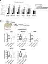
Figure 5. DNMT inhibitors reactivate striatal gene expression in mutant Htt-expressing primary neurons and R6/2 HD mouse brain in vivo.
(A) DIV 5 mouse primary striatal neurons were infected with lentivirus expressing Htt-25Q or Htt-72Q exon1 fragment; 5 days later, RNA was prepared and subjected to qRT-PCR analysis. β-actin and Hprt were used as reference genes. Decitabine restored the expression of downregulated genes in mutant Htt-expressing striatal neurons (ANOVA, *P < 0.05, n = 3 compared to Htt-72Q plus vehicle. Similar results were observed when Htt-72Q-expressing neurons were treated with FdCyd (data not shown). Data are presented as mean + SEM. (B) (Top) Procedure for the treatment of mice with FdCyd. A mini-osmotic pump containing FdCyd (0.1 mM in saline) was implanted subcutaneously on the back of mice at 6 weeks of age, and the drug was infused into the right ventricle through a stereotactically placed catheter. One week later, the striatum was dissected for qRT-PCR analysis. ICV, intracerebroventricular. (Bottom) FdCyd was delivered into R6/2 or WT mouse brain by icv infusion at 6 weeks of age. Saline was used as control. One week after drug infusion was initiated, RNA was extracted from the striatum and subjected to qRT-PCR analysis. β-actin was used as a reference gene. Levels of Drd2, Ppp1r1b, Rasd2, and Adora2a mRNA were restored in R6/2 mice after FdCyd treatment. FdCyd treatment showed a trend towards increasing Penk RNA in R6/2 striatum. (ANOVA, *P < 0.005 compared to WT–saline, #P < 0.05 compared to R6/2–saline, n = 7–9 mice per group). The vertical bars represent the range of values.
Figure 6
Figure 6. A model for the role of DNA methylation in HD neurodegeneration.
Inhibition of DNMTs in HD neurons by pharmacological inhibitors (decitabine or FdCyd) or RNAi blocks mutant Htt-induced neurotoxicity as well as transcriptional repression of key genes, such as Bdnf, Drd2, Ppp1r1b, and Adora2a. The DNA methylation pathway may thus play an important role in HD neurodegeneration.

Similar articles

Cited by

References

    1. Walker F. O. Huntington’s disease. Lancet 369, 218–228 (2007). - PubMed
    1. Ross C. A. et al.. Huntington disease: natural history, biomarkers and prospects for therapeutics. Nature reviews. Neurology 10, 204–216 (2014). - PubMed
    1. The Huntington’s Disease Collaborative Research Group. A novel gene containing a trinucleotide repeat that is expanded and unstable on Huntington’s disease chromosomes. Cell 72, 971–983 (1993). - PubMed
    1. Sugars K. L. & Rubinsztein D. C. Transcriptional abnormalities in Huntington disease. Trends Genet 19, 233–238 (2003). - PubMed
    1. Cha J. H. Transcriptional signatures in Huntington’s disease. Prog Neurobiol 83, 228–248 (2007). - PMC - PubMed

Publication types

MeSH terms