Review
. 2016 Oct;38(10):969-76.
doi: 10.1002/bies.201600067. Epub 2016 Aug 12.

In search of a periodic table of the neurons: Axonal-dendritic circuitry as the organizing principle: Patterns of axons and dendrites within distinct anatomical parcels provide the blueprint for circuit-based neuronal classification

Affiliations
Review

In search of a periodic table of the neurons: Axonal-dendritic circuitry as the organizing principle: Patterns of axons and dendrites within distinct anatomical parcels provide the blueprint for circuit-based neuronal classification

Giorgio A Ascoli et al. Bioessays. 2016 Oct.

Abstract

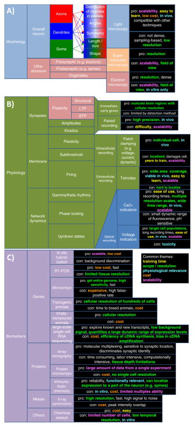
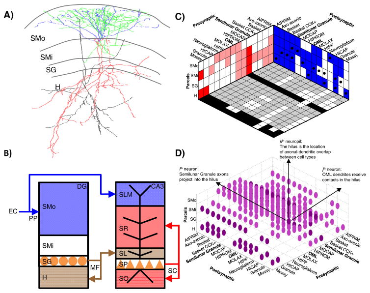
No one knows yet how to organize, in a simple yet predictive form, the knowledge concerning the anatomical, biophysical, and molecular properties of neurons that are accumulating in thousands of publications every year. The situation is not dissimilar to the state of Chemistry prior to Mendeleev's tabulation of the elements. We propose that the patterns of presence or absence of axons and dendrites within known anatomical parcels may serve as the key principle to define neuron types. Just as the positions of the elements in the periodic table indicate their potential to combine into molecules, axonal and dendritic distributions provide the blueprint for network connectivity. Furthermore, among the features commonly employed to describe neurons, morphology is considerably robust to experimental conditions. At the same time, this core classification scheme is suitable for aggregating biochemical, physiological, and synaptic information.

Keywords: axons; circuits; classification; dendrites; neurons.


Conflict of interest statement

None of the authors has a conflict of interest to declare.

Figures

Figure 1
Figure 1
The three main experimental approaches to characterize neuron types. A: Morphology. B: Electrophysiology. C: Molecular biomarkers. Each approach is suitable to investigate a subset of general properties and specific features based on different techniques. The advantages and disadvantages of every technique are color-coded along themes that recur across dimensions: invasiveness, cost, scalability, training time, coverage scope, and resolution. D: The GABAergic interneurons of hippocampal area CA1 can be grouped on the basis of selected properties across three dimensions in a highly simplified binary classification scheme. In this example, neuron types are divided along the vertical axis based on parvalbumin (PV) expression (one of many molecular biomarkers), along the depth axis based on firing threshold potential (Vthresh; one of many electrophysiological variables), and along the horizontal axis as to whether they can receive perforant path (PP) input (one of many morphological factors).
Figure 1
Figure 1
The three main experimental approaches to characterize neuron types. A: Morphology. B: Electrophysiology. C: Molecular biomarkers. Each approach is suitable to investigate a subset of general properties and specific features based on different techniques. The advantages and disadvantages of every technique are color-coded along themes that recur across dimensions: invasiveness, cost, scalability, training time, coverage scope, and resolution. D: The GABAergic interneurons of hippocampal area CA1 can be grouped on the basis of selected properties across three dimensions in a highly simplified binary classification scheme. In this example, neuron types are divided along the vertical axis based on parvalbumin (PV) expression (one of many molecular biomarkers), along the depth axis based on firing threshold potential (Vthresh; one of many electrophysiological variables), and along the horizontal axis as to whether they can receive perforant path (PP) input (one of many morphological factors).
Figure 2
Figure 2
Axonal-dendritic patterns and anatomical regions: two sides of the same coin. A: Morphological reconstructions (from NeuroMorpho.Org [50]) of a rat dentate gyrus semilunar granule cell (dendrites in blue, axons in red; NMO_10035 from Ref. [28]) and of an OML cell (dendrites in black, axons in green; NMO_00178 from Ref. [31]). B: The layers of the dentate gyrus (DG) and hippocampal area CA3 are defined by the somatic locations and axonal terminals of principal neurons. The perforant path (PP) axonal terminals of entorhinal layer 2 spiny stellate cells delineate the outer molecular layer in DG and lacunosum-moleculare in CA3. The mossy fiber (MF) axonal terminals of DG granule cells define the hilus in DG and the lucidum layer in CA3. The recurrent and commissural terminals of CA3 pyramidal cells demarcate the oriens and radiatum layers in CA3. Abbreviations: SMo (outer stratum moleculare), SMi (inner stratum moleculare), SG (stratum granulare), H (hilus), SLM (stratum lacunosum-moleculare), SR (stratum radiatum), SL (stratum lucidum), SP (stratum pyramidale), and SO (stratum oriens). C: A periodic table of neuromorphological patterns in the dentate gyrus. The two walls of the box represent the pre-synaptic axonal (red) and post-synaptic dendritic (blue) binary distributions of every neuron type (listed horizontally) across all neuropils (listed vertically). The box floor represents the predicted circuitry, in which black and gray squares correspond to potential excitatory and inhibitory connections, respectively, and white squares indicate absence of synapses. Black dots on the blue wall indicate the location of the somata for the various neuron types. D: A “raindrop cloud” representation of the neuropil tensor, where the ijkth entry represents a potential synapse formed by the axon of the ith neuron onto the dendrite of the jth neuron in the kth neuropil.
Figure 3
Figure 3
Periodic tables and potential circuit connectivity (encoding as in Figure 2, C and D). A: Neuron types from the cat primary visual neocortex: layer 2/3 pyramidal cells (p2/3); layer 2/3 basket cells (b2/3); layer 2/3 double bouquet cells (db2/3); layer 4 spiny stellate cells with axon in layer 4 (ss4 (L4)); layer 4 spiny stellate cells with axon in layer 2/3 (ss4 (L2/3)); layer 4 pyramidal cells (p4); layer 4 basket cells (b4); layer 5 pyramidal cells with axon in layer 2/3 (p5 (L2/3)); layer 5 pyramidal cells with axon in layers 5 and 6 (p5 (L5/6)); layer 5 basket cells (b5); layer 6 pyramidal cells with axon in layer 4 (p6 (L4)); layer 6 pyramidal cells with axon in layers 5 and 6 (p6 (L5/6)). B: Potential connectivity of the neurons represented in panel A. C: Cholinergic (excitatory) and GABAergic (inhibitory) neuron types from the fly brain (see Supplementary Information), with birth day indicated. D: Potential connectivity of the neurons represented in panel A.

References

    1. Huang ZJ. Toward a genetic dissection of cortical circuits in the mouse. Neuron. 2014;83:1284–302. - PMC - PubMed
    1. Denk W, Briggman KL, Helmstaedter M. Structural neurobiology: missing link to a mechanistic understanding of neural computation. Nat Rev Neurosci. 2012;13:351–8. - PubMed
    1. Lodato S, Arlotta P. Generating neuronal diversity in the mammalian cerebral cortex. Annu Rev Cell Dev Biol. 2015;31:699–720. - PMC - PubMed
    1. Sharpee TO. Toward functional classification of neuronal types. Neuron. 2014;83:1329–34. - PMC - PubMed
    1. Kepecs A, Fishell G. Interneuron cell types are fit to function. Nature. 2014;505:318–26. - PMC - PubMed

Publication types

LinkOut - more resources