Chronic exposure to haloperidol and olanzapine leads to common and divergent shape changes in the rat hippocampus in the absence of grey-matter volume loss
- PMID: 27516217
- PMCID: PMC5108303
- DOI: 10.1017/S0033291716001768
Chronic exposure to haloperidol and olanzapine leads to common and divergent shape changes in the rat hippocampus in the absence of grey-matter volume loss
Abstract
Background: One of the most consistently reported brain abnormalities in schizophrenia (SCZ) is decreased volume and shape deformation of the hippocampus. However, the potential contribution of chronic antipsychotic medication exposure to these phenomena remains unclear.
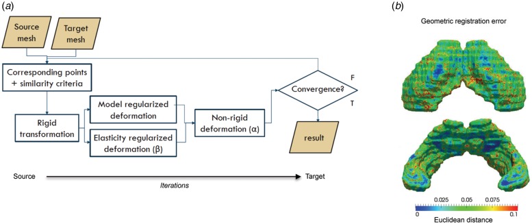
Method: We examined the effect of chronic exposure (8 weeks) to clinically relevant doses of either haloperidol (HAL) or olanzapine (OLZ) on adult rat hippocampal volume and shape using ex vivo structural MRI with the brain retained inside the cranium to prevent distortions due to dissection, followed by tensor-based morphometry (TBM) and elastic surface-based shape deformation analysis. The volume of the hippocampus was also measured post-mortem from brain tissue sections in each group.
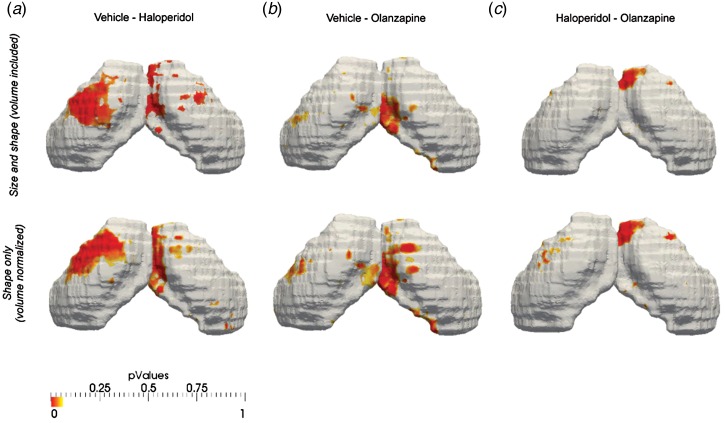
Results: Chronic exposure to either HAL or OLZ had no effect on the volume of the hippocampus, even at exploratory thresholds, which was confirmed post-mortem. In contrast, shape deformation analysis revealed that chronic HAL and OLZ exposure lead to both common and divergent shape deformations (q = 0.05, FDR-corrected) in the rat hippocampus. In particular, in the dorsal hippocampus, HAL exposure led to inward shape deformation, whereas OLZ exposure led to outward shape deformation. Interestingly, outward shape deformations that were common to both drugs occurred in the ventral hippocampus. These effects remained significant after controlling for hippocampal volume suggesting true shape changes.
Conclusions: Chronic exposure to either HAL or OLZ leads to both common and divergent effects on rat hippocampal shape in the absence of volume change. The implications of these findings for the clinic are discussed.
Keywords: antipsychotic; hippocampus; magnetic resonance imaging; schizophrenia; shape; volume.
Figures


Similar articles
-
Olanzapine counteracts reduction of brain-derived neurotrophic factor and TrkB receptors in rat hippocampus produced by haloperidol.Neurosci Lett. 2004 Feb 12;356(2):135-9. doi: 10.1016/j.neulet.2003.10.079. Neurosci Lett. 2004. PMID: 14746882
-
Microglial activation in the rat brain following chronic antipsychotic treatment at clinically relevant doses.Eur Neuropsychopharmacol. 2015 Nov;25(11):2098-107. doi: 10.1016/j.euroneuro.2015.08.004. Epub 2015 Aug 17. Eur Neuropsychopharmacol. 2015. PMID: 26321204
-
Differential effects of haloperidol and olanzapine on the expression of erythropoietin and its receptor in rat hippocampus and striatum.J Neurochem. 2006 Sep;98(5):1411-22. doi: 10.1111/j.1471-4159.2006.04057.x. J Neurochem. 2006. PMID: 16923156
-
Modulation of nerve growth factor and choline acetyltransferase expression in rat hippocampus after chronic exposure to haloperidol, risperidone, and olanzapine.Psychopharmacology (Berl). 2004 Apr;172(4):365-74. doi: 10.1007/s00213-003-1669-6. Epub 2003 Nov 28. Psychopharmacology (Berl). 2004. PMID: 14647958
-
Prior haloperidol, but not olanzapine, exposure augments the pursuit of reward cues: implications for substance abuse in schizophrenia.Schizophr Bull. 2013 May;39(3):692-702. doi: 10.1093/schbul/sbs077. Epub 2012 Aug 27. Schizophr Bull. 2013. PMID: 22927669 Free PMC article.
Cited by
-
Normalizing the Abnormal: Do Antipsychotic Drugs Push the Cortex Into an Unsustainable Metabolic Envelope?Schizophr Bull. 2020 Apr 10;46(3):484-495. doi: 10.1093/schbul/sbz119. Schizophr Bull. 2020. PMID: 31755955 Free PMC article. Review.
-
Synaptic density marker SV2A is reduced in schizophrenia patients and unaffected by antipsychotics in rats.Nat Commun. 2020 Jan 14;11(1):246. doi: 10.1038/s41467-019-14122-0. Nat Commun. 2020. PMID: 31937764 Free PMC article.
-
Role of D3 dopamine receptors in modulating neuroanatomical changes in response to antipsychotic administration.Sci Rep. 2019 May 24;9(1):7850. doi: 10.1038/s41598-019-43955-4. Sci Rep. 2019. PMID: 31127135 Free PMC article.
-
Neuroinflammation in schizophrenia: meta-analysis of in vivo microglial imaging studies.Psychol Med. 2019 Oct;49(13):2186-2196. doi: 10.1017/S0033291718003057. Epub 2018 Oct 25. Psychol Med. 2019. PMID: 30355368 Free PMC article. Review.
-
The Effects of Antipsychotics on the Synaptic Plasticity Gene Homer1a Depend on a Combination of Their Receptor Profile, Dose, Duration of Treatment, and Brain Regions Targeted.Int J Mol Sci. 2020 Aug 3;21(15):5555. doi: 10.3390/ijms21155555. Int J Mol Sci. 2020. PMID: 32756473 Free PMC article.
References
-
- Altshuler LL, Bartzokis G, Grieder T, Curran J, Jimenez T, Leight K, Wilkins J, Gerner R, Mintz J (2000). An MRI study of temporal lobe structures in men with bipolar disorder or schizophrenia. Biological Psychiatry 48, 147–162. - PubMed
-
- Arango C, Breier A, McMahon R, Carpenter WT Jr., Buchanan RW (2003). The relationship of clozapine and haloperidol treatment response to prefrontal, hippocampal, and caudate brain volumes. American Journal of Psychiatry 160, 1421–1427. - PubMed
-
- Bannerman DM, Rawlins JN, McHugh SB, Deacon RM, Yee BK, Bast T, Zhang WN, Pothuizen HH, Feldon J (2004). Regional dissociations within the hippocampus – memory and anxiety. Neuroscience Biobehavioural Reviews 28, 273–283. - PubMed
MeSH terms
Substances
Grants and funding
LinkOut - more resources
Full Text Sources
Other Literature Sources