Review
. 2016 Sep;27(9):2564-75.
doi: 10.1681/ASN.2016020177. Epub 2016 Aug 11.

Of Inflammasomes and Alarmins: IL-1β and IL-1α in Kidney Disease

Affiliations
Review

Of Inflammasomes and Alarmins: IL-1β and IL-1α in Kidney Disease

Hans-Joachim Anders. J Am Soc Nephrol. 2016 Sep.

Abstract

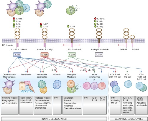
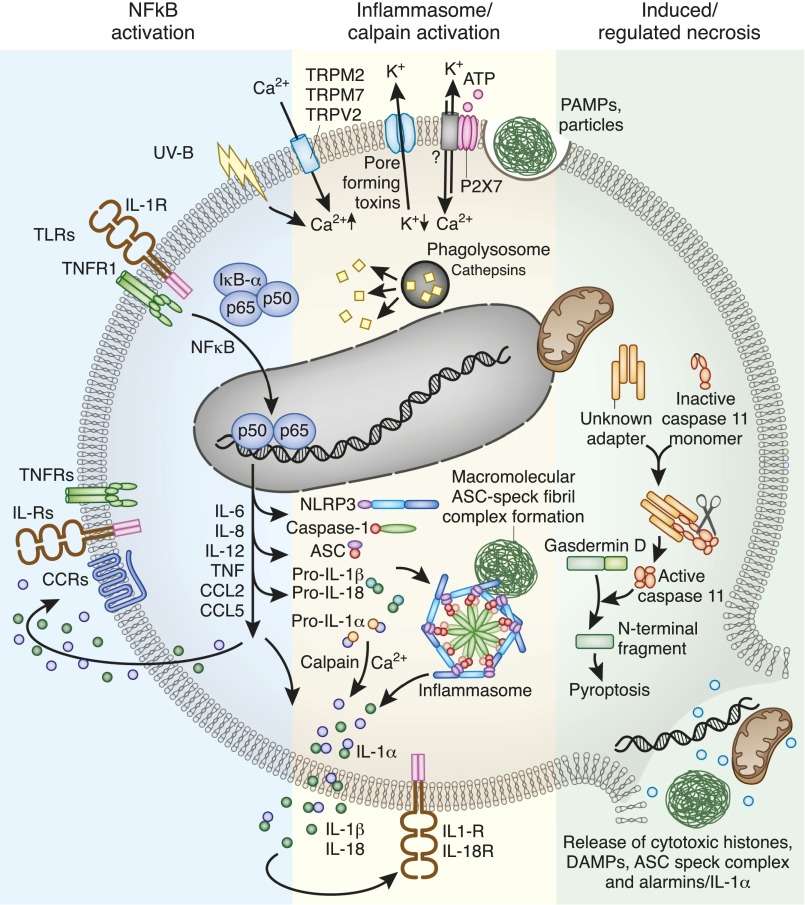
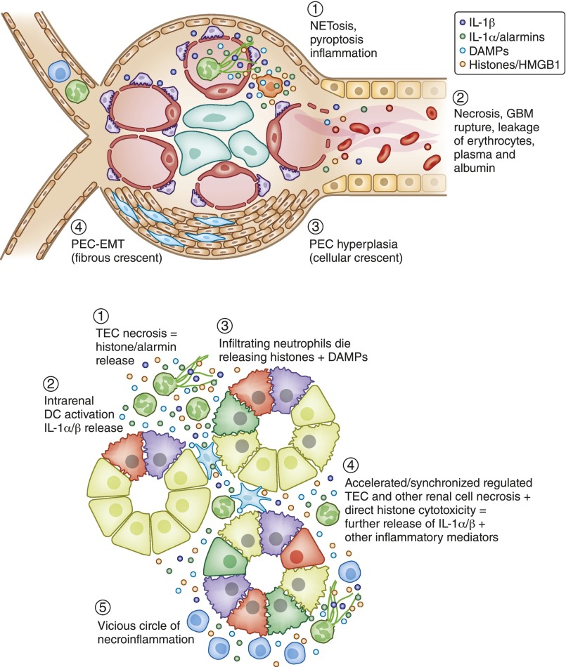
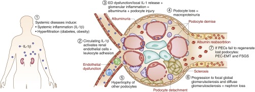
Kidney injury implies danger signaling and a response by the immune system. The inflammasome is a central danger recognition platform that triggers local and systemic inflammation. In immune cells, inflammasome activation causes the release of mature IL-1β and of the alarmin IL-1α Dying cells release IL-1α also, independently of the inflammasome. Both IL-1α and IL-1β ligate the same IL-1 receptor (IL-1R) that is present on nearly all cells inside and outside the kidney, further amplifying cytokine and chemokine release. Thus, the inflammasome-IL-1α/IL-β-IL-1R system is a central element of kidney inflammation and the systemic consequences. Seminal discoveries of recent years have expanded this central paradigm of inflammation. This review gives an overview of arising concepts of inflammasome and IL-1α/β regulation in renal cells and in experimental kidney disease models. There is a pipeline of compounds that can interfere with the inflammasome-IL-1α/IL-β-IL-1R system, ranging from recently described small molecule inhibitors of NLRP3, a component of the inflammasome complex, to regulatory agency-approved IL-1-neutralizing biologic drugs. Based on strong theoretic and experimental rationale, the potential therapeutic benefits of using such compounds to block the inflammasome-IL-1α/IL-β-IL-1R system in kidney disease should be further explored.

Keywords: acute kidney injury; chronic kidney disease; glomerulonephritis; inflammation; innate immunity.


Figures

Figure 1.
Figure 1.
The families of IL-1 cytokines and cytokine receptors activate innate and adaptive immunity. Activated or dying cells release all sorts of cytokines of the IL-1 family that specifically interact with several transmembrane surface receptors present on most cell types of the body. Some induce cell activation (IL-1R, IL-18R, IL-36R), others inhibit cell activation (IL-33R, TIGIRR, SIGIRR). This way the family elicits numerous regulatory effects on renal cells, immune cells of the innate and adaptive immune system, either activating or inhibiting their respective cell type–specific functions. AG, antigen; G-CSF, granulocyte colony-stimulating factor; MФ, macrophage; NETs, neutrophil extracellular traps; NK cell, natural killer cell; TIR domain, Toll/interleukin-1 receptor (TIR) homology domain.
Figure 2.
Figure 2.
Inflammasome activation in dendritic cells and macrophages involves numerous elements. Dendritic cells first need to induce the expression of the inflammasome components and of pro–IL-1α and pro–IL-1β. This can occur via cytokine receptors or Toll-like receptors (TLRs). Activation of inflammasome assembly can occur upon numerous intracellular danger signals such as mitochondrial reactive oxygen species release, lysosomal protease leakage, and potassium efflux or calcium influx. The multiprotein inflammasome complex forms a wheel-like structure to trigger caspase-1–driven IL-1β (and IL-18) enzymatic activation. Inflammasome fibrils grow in size to a single macromolecular complex called ASC speck. Calcium activates calpain, which cleaves pro–IL-1α to IL-1α, but in contrast to pro–IL-1β, IL-1α is already biologically active, and hence an alarmin. IL-1α, IL-1β, and IL-18 together with other NF-κB–dependent cytokines and chemokines activate cytokine and chemokine receptors in an autocrine, paracrine, or systemic manner. Noncanonical inflammasome signaling, e.g., triggered by cytosolic LPS, involves caspase-11 (mice) and caspase 4/5 (humans), which cleave gasdermin D. The cleaving product activates NLRP3. Noncanonical inflammasome signaling is a recognized trigger for pyroptosis, an immunogenic form of cell death that leads to the release of numerous intracellular components that have the capacity to activate a plethora of pattern recognition receptors and close the vicious cycle of necroinflammation. CCL, CC chemokine ligand; CCR, CC-chemokine receptor.
Figure 3.
Figure 3.
The inflammasome/IL-1 system contributes to renal necroinflammation. Necroinflammation can occur in the glomerulus (A) or the tubulointerstitial compartment (B). The primary event can be intravascular NETosis, as in ANCA vasculitis, or renal cell necrosis, such as in ischemic tubular injury. Histone, DAMP, and alarmin (IL-1α) released from dying cells activate the NLRP3 inflammasome (IL-1α/β release) and induce IL-1R signaling, which implies local inflammation. Especially histones also kill other cells, resulting in a crescendo of tissue inflammation and necrosis, i.e., necroinflammation. The consequences in the glomerulus and the tubules are illustrated. Both lead to a drop out of nephrons and the clinical syndrome of AKI.
Figure 4.
Figure 4.
The inflammasome/IL-1 system promotes CKD. CKD is devoid of tissue necrosis and currently there is little evidence for renal cell necroptosis or other forms of regulated cell necrosis being involved in CKD progression. There are data supporting a role of the NLRP3 inflammasome and IL-1 in CKD but the mechanisms are unclear. Most likely systemic release of IL-1, e.g., in diabetes or systemic lupus, contributes to systemic endothelial dysfunction (ED), a process also promoting leukocyte adhesion and vascular leakage (microalbuminuria) in the kidney. Furthermore, intrarenal inflammasome activation in immune cells, and eventually also in renal parenchymal cells, may contribute to local inflammation, cell stress, and cell loss. For example, podocyte detachment promotes albuminuria, hypertrophy of the remaining podocytes, FSGS, and subsequently first focal-global and later diffuse glomerulosclerosis-related nephron loss. In this process the inflammasome components NLRP3 and ASC may contribute to SMAD phosphorylation downstream of TGFR signaling during epithelial-mesenchymal transition (EMT) of parietal epithelial cells (PEC), and induce extracellular matrix production by tubular epithelial cells (not depicted). Whether the same also occurs in interstitial fibroblasts is currently unknown (not depicted). A role of NLRP3 and ASC has also been reported on systemic autoimmunity where that assures the immunosuppressive effect of TGFR signaling on immune cells (not depicted).

References

    1. Anders HJ, Muruve DA: The inflammasomes in kidney disease. J Am Soc Nephrol 22: 1007–1018, 2011 - PubMed
    1. Auron PE, Webb AC, Rosenwasser LJ, Mucci SF, Rich A, Wolff SM, Dinarello CA: Nucleotide sequence of human monocyte interleukin 1 precursor cDNA. Proc Natl Acad Sci U S A 81: 7907–7911, 1984 - PMC - PubMed
    1. Lomedico PT, Gubler U, Hellmann CP, Dukovich M, Giri JG, Pan YC, Collier K, Semionow R, Chua AO, Mizel SB: Cloning and expression of murine interleukin-1 cDNA in Escherichia coli. Nature 312: 458–462, 1984 - PubMed
    1. Garlanda C, Dinarello CA, Mantovani A: The interleukin-1 family: back to the future. Immunity 39: 1003–1018, 2013 - PMC - PubMed
    1. Netea MG, van de Veerdonk FL, van der Meer JW, Dinarello CA, Joosten LA: Inflammasome-independent regulation of IL-1-family cytokines. Annu Rev Immunol 33: 49–77, 2015 - PubMed