. 2016 Oct;172(2):980-1002.
doi: 10.1104/pp.16.00690. Epub 2016 Aug 11.

Exocyst SEC3 and Phosphoinositides Define Sites of Exocytosis in Pollen Tube Initiation and Growth

Affiliations

Exocyst SEC3 and Phosphoinositides Define Sites of Exocytosis in Pollen Tube Initiation and Growth

Daria Bloch et al. Plant Physiol. 2016 Oct.

Abstract

Polarized exocytosis is critical for pollen tube growth, but its localization and function are still under debate. The exocyst vesicle-tethering complex functions in polarized exocytosis. Here, we show that a sec3a exocyst subunit null mutant cannot be transmitted through the male gametophyte due to a defect in pollen tube growth. The green fluorescent protein (GFP)-SEC3a fusion protein is functional and accumulates at or proximal to the pollen tube tip plasma membrane. Partial complementation of sec3a resulted in the development of pollen with multiple tips, indicating that SEC3 is required to determine the site of pollen germination pore formation. Time-lapse imaging demonstrated that SEC3a and SEC8 were highly dynamic and that SEC3a localization on the apical plasma membrane predicts the direction of growth. At the tip, polar SEC3a domains coincided with cell wall deposition. Labeling of GFP-SEC3a-expressing pollen with the endocytic marker FM4-64 revealed the presence of subdomains on the apical membrane characterized by extensive exocytosis. In steady-state growing tobacco (Nicotiana tabacum) pollen tubes, SEC3a displayed amino-terminal Pleckstrin homology-like domain (SEC3a-N)-dependent subapical membrane localization. In agreement, SEC3a-N interacted with phosphoinositides in vitro and colocalized with a phosphatidylinositol 4,5-bisphosphate (PIP2) marker in pollen tubes. Correspondingly, molecular dynamics simulations indicated that SEC3a-N associates with the membrane by interacting with PIP2 However, the interaction with PIP2 is not required for polar localization and the function of SEC3a in Arabidopsis (Arabidopsis thaliana). Taken together, our findings indicate that SEC3a is a critical determinant of polar exocytosis during tip growth and suggest differential regulation of the exocytotic machinery depending on pollen tube growth modes.


Figures

Figure 1.
Figure 1.
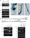
Expression pattern of the SEC3a and SEC3b paralogs and genotyping of sec3a-1 and sec3b-1 mutants. A, RT-PCR showing the expression of SEC3a and SEC3b in seedlings at 6 and 14 d after germination (DAG), different mature plant organs, closed flowers (Fc), ovules, anthers (fully developed from closed flowers), and in vitro-germinated pollen. B to D, Expression pattern of SEC3a (PSEC3a:GUS) in seedlings. Four independent lines were used for analysis. B, A 2-DAG seedling showing the highest expression levels in cotyledons and primary root tips (arrowheads). C, A primary root of a 2-DAG seedling. dfz, Differentiation zone; dz, division zone; ez, elongation zone. The arrowhead marks expression in root hairs. D, A root with an emerging lateral root primordium. Bars = 50 µm. E, Schematic representation of SEC3a and SEC3b gene exon-intron organization. Gene-specific primers used for genotyping and determining the T-DNA insert locations are specified by arrows. Numbers indicate nucleotide positions from the initiation ATG codon. The T-DNA inserts at positions 6,673 in SEC3a and 4,529 in SEC3b are highlighted by arrowheads. F, RT-PCR showing the expression of SEC3a and SEC3b in flowers of the indicated genotypes. Fo, Open flower. A genomic DNA control was included to display primer efficiency. G, SEC3a and SEC3b expression in sec3a-1/LAT52:SEC3a complemented plants. Note that in sec3a-1−/−/LAT52:SEC3a homozygous lines, expression of the SEC3a coding sequence (CDS) indicates that the SEC3a transgene construct was in the sporophyte (for primer details, see Supplemental Fig. S1).
Figure 2.
Figure 2.
Tetrad analysis of sec3a-1 heterozygotes. A, In vitro-germinated tetrads from self-crosses of qrt1−/− and qrt1−/−/sec3a-1+/− plants. The chart shows the percentage from total quartets with the indicated number of germinated pollen grains in each tetrad (between zero and four) compared with the total tetrads scored for each genotype. Numbers above the bars represent actual numbers of tetrads that were counted. B, Aniline Blue staining of wild-type stigma 24 h after pollination with either qrt1−/− or qrt1−/−/sec3a-1+/− tetrads. Asterisks mark germinated pollen grains in tetrads as revealed by the staining. The chart shows in vivo germination analysis of qrt1−/− and qrt1−/−/sec3a-1+/− tetrads germinated on Col-0 stigma. The number of pollen tubes producing cells in each quartet (between one and four) was counted using Aniline Blue staining. Bars = 50 µm.
Figure 3.
Figure 3.
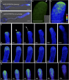
Localization of GFP-SEC3a in growing Arabidopsis pollen tubes. A, In vitro-germinated Arabidopsis pollen stably expressing LAT52:GFP-SEC3a. B, Apex of the LAT52:GFP-SEC3a pollen tube. The arrowhead marks the membrane localization at the tip. C, The same apex shown in B with GFP signal converted into an intensity scale. D, A nongrowing pollen tube stably expressing LAT52:GFP-SEC3a. Note the absence of the protein from the tip. E, Selected time-lapse images showing the localization of GFP-SEC3a in a growing pollen tube. Note that apical localization of GFP-SEC3a is dynamic and the protein accumulates at the future direction of tube growth (arrowhead). The arrow represents the direction of growth as predicted by GFP-SEC3a localization. F, Localization of GFP-SEC3a in a nongrowing pollen tube. The intensity of the GFP signal decreases with time due to high magnification and frequent imaging, and the last frame (210 s) shows the final direction of tube growth after 3.5 min. Note the cone-shape accumulation of fluorescent signals in growing pollen tubes and their absence in nongrowing tubes. G, Time-lapse images showing the localization of GFP-SEC8 in a growing pollen tube of an in vitro-germinated Arabidopsis pollen, stably expressing PSEC8:GFP-SEC8 in a sec8-1 mutant background. In A, C, and D to G, GFP fluorescence is represented as an intensity color scale. Bars = 10 µm in A and D and 5 µm in B, C, and E to G.
Figure 4.
Figure 4.
Labeling of GFP-SEC3a Arabidopsis pollen tubes with PI. GFP-SEC3a Arabidopsis pollen tubes were stained with 10 µm PI. A, Apex of a pollen tube showing the localization of GFP-SEC3a and PI with intensity scale for each channel. Asterisks mark positions on the intensity plot in B. B, Intensity plot of GFP and PI signals across the plasma membrane/cell wall of pollen apex (including flanks). Asterisks mark positions on the corresponding image in A. C, Time-lapse images of a growing pollen tube. Note that GFP-SEC3a decorates the PM beneath the strongest PI signal at the tip. D, Enlarged images of the pollen tip showing thick cell wall at the tip (arrowheads) and GFP-SEC3a signal decorating the PM at this region. a.u., Arbitrary units. Bars = 5 µm.
Figure 5.
Figure 5.
Labeling of GFP-SEC3a Arabidopsis pollen tubes with FM4-64. GFP-SEC3a Arabidopsis pollen tubes were labeled with 1 µm FM4-64 for 3 min (A–C) or 20 min (D–F) and then subjected to imaging. A and D, Apex of the pollen tube showing the localization of GFP-SEC3a and FM4-64 with intensity scale for each channel. Asterisks mark positions on the intensity plots in B and E. The arrowhead in D points to the region with high GFP-SEC3a and low FM4-64 signals. B and E, Intensity plots of GFP and FM4-64 signal across the PM of the pollen apex (including flanks). Asterisks mark positions on the corresponding images in A and D. C and F, Time-lapse images showing the localization of GFP-SEC3a and FM4-64 in growing pollen tubes. In F, the white arrows mark the direction of pollen tube elongation as predicted from tip geometry, and the yellow arrow indicates the actual growth direction. Note that the position of high GFP and low FM4-64 signals correlates with growth direction. a.u., Arbitrary units. Bars = 5 µm.
Figure 6.
Figure 6.
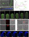
Expression of GFP-SEC3a in the sec3a-1 homozygous background results in the development of pollen with multiple tips. A, Germinating sec3a-1 pollen expressing GFP-SEC3a. The multiple tips are marked by numbers. B, In pollen tubes with one tip, GFP-SEC3a concentrates at the apex (GFP fluorescence is shown as an intensity color scale). C, Selected time-lapse images of sec3a-1/GFP-SEC3a pollen labeled with 1 µm FM4-64 for 20 min. Note that GFP-SECa effectively accumulates at the apical plasma membrane. D, Intensity plot of GFP and FM4-64 signal across the PM of pollen apex (including flanks). Asterisks mark positions on the corresponding image in C. E to H, Immunolabeling of deesterified pectins (E and F) and esterified pectins (G and H) with LM19 and LM20 monoclonal antibodies, respectively. E and G, Nontransgenic Col-0 pollen. F and H, sec3a-1/GFP-SEC3a pollen. The white rectangles in E and G highlight the enlarged regions shown in the middle image of each panel. The black arrows highlight germinated pollen tubes in F and the actively growing pollen tubes in H. The yellow arrows in H highlight the arrested small pollen tubes. I, Localization of GFP-SEC3a in multiple-tip sec3a-1/GFP-SEC3a pollen. The white arrow denotes the growing pollen tube, and the yellow arrow indicates the arrested pollen tube. See also Supplemental Movies S4 and S5. a.u., Arbitrary units. Bars = 20 µm in A, B, E, and G, 10 µm in F, H, and I, and 5 µm in C.
Figure 7.
Figure 7.
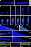
The N-terminal PH domain of Arabidopsis SEC3a colocalizes with PIP2 and is responsible for the localization of SEC3a at the plasma membrane in tobacco pollen tubes. A, Localization of full-length YFP-SEC3a transiently expressed in tobacco pollen tubes under the control of the LAT52 promoter. B and C, Selected time-lapse images showing the localization of LAT52:YFP-SEC3a in steady-growing (B) and oscillating (C) pollen tubes. D and E, Kymograph analysis of growth and apical YFP-SEC3a fluorescence in steady-growing (D) and oscillating (E) pollen tubes. Arrowheads in C and E point to dynamic apical localization of SEC3a in oscillating pollen tubes. F and G, Localization (F) and kymograph analysis (G) of the SEC3a N-terminal domain (LAT52:YFP-SEC3a-N). H and I, Localization (H) and kymograph analysis (I) of SEC3a lacking its N-terminal domain (LAT52:YFP-SEC3a-ΔN). J, Colocalization of YFP-SEC3a-N with the PIP2 marker mRFP1-PHPLCδ1. In A to I, YFP fluorescence is represented as an intensity color scale. PLC, Phospholipase C. Bars = 5 µm.
Figure 8.
Figure 8.
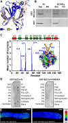
SEC3a-N binds phosphoinositides via four positively charged residues that mediate its association with the plasma membrane in tobacco pollen tubes. A, Comparison of the final relaxed SEC3a-N homology model (At) with the yeast template structure (Sc). B, Liposome-binding assay of GST-SEC3a-N. PIP2 binding was determined using 200-nm vesicles containing 5% PIP2:95% PC or PC alone. After incubation of GST-SEC3a-N with the vesicles, they were recovered by ultracentrifugation, and protein bound (Pel) was analyzed by SDS-PAGE. As a negative control, GST alone was used. Representative results from three independent experiments are shown. Sup, Supernatant. C, Analysis of MD simulations displaying polar contacts of SEC3a-N with a PIP2 molecule. The bars represent average numbers of polar contacts through the last 8 μs of the simulation. Polar contacts were defined as the number of PIP2 head group atoms within 8 Å of protein atoms. The inset image represents a coarse-grained model of SEC3a-N coordinating the PIP2 molecule. The interacting residues are highlighted. D, Lipid-binding properties of wild-type GST-SEC3a-N and mutated GST-SEC3a-N KRKR/A recombinant proteins, as determined using a protein-lipid overlay assay. DAG, Diacylglycerol; PA, phosphatidic acid; PE, phosphatidylethanolamine; PG, phosphatidylglycerol; PI4P, phosphatidylinositol 4-phosphate; PI4,5P2, phosphatidylinositol 4,5-bisphosphate; PI3,4,5P3, phosphatidylinositol 3,4,5-trisphosphate; PS, phosphatidylserine; TAG, triacylglycerol. E, Localization of wild-type YFP-SEC3a-N and the mutated variant YFP-SEC3a-N KRKR/A in tobacco pollen tubes. YFP fluorescence is represented as an intensity color scale. Bars = 5 µm.
Figure 9.
Figure 9.
The N-terminal PH domain of SEC3a is not required for its accumulation at the tip of growing Arabidopsis pollen tubes. A to C, In vitro-germinated Arabidopsis pollen tubes stably expressing GFP-SEC3a (A) GFP-SEC3a KRKR/A (B), and GFP-SEC3-ΔN (C) under the regulation of the LAT52 promoter. D, Selected time-lapse images of growing GFP-SEC3a, GFP-SEC3a KRKR/A, and GFP-SEC3a-ΔN Arabidopsis pollen tubes. GFP fluorescence is represented as an intensity color scale. Bars = 10 µm in A to C and 5 µm in D.
Figure 10.
Figure 10.
SEC3a function and localization in pollen tube tip growth. A, SEC3a localization at the tip PM is dynamic and defines the direction of pollen tube growth. Secretory vesicles, presumably loaded with pectins, are delivered to the region on the tip PM marked by SEC3a, resulting in the local deposition of pectins. B, In elongating pollen tubes, the PM at the tip is characterized by a region with high GFP-SEC3a and low FM4-64 signals, which could be considered a polar domain of focused and extensive exocytosis. In nonelongating pollen tubes, GFP-SEC3 signal on the PM is homogenously distributed across the tip and shank PM. As a consequence, a polar domain for exocytosis is not established and the tubes grow isotropically. C, In tobacco, SEC3a PM localization depends on the mode of pollen tube growth. During steady growth, SEC3a localizes to a subapical region and requires PIP2 for the association with membrane. In oscillatory growing pollen tubes, SEC3a association with the PM at the tip is dynamic, as in Arabidopsis. The difference in SEC3a localization between these two growing modes likely reflects the actual site for the delivery of secretory vesicles.

References

    1. Baek K, Knödler A, Lee SH, Zhang X, Orlando K, Zhang J, Foskett TJ, Guo W, Dominguez R (2010) Structure-function study of the N-terminal domain of exocyst subunit Sec3. J Biol Chem 285: 10424–10433 - PMC - PubMed
    1. Boavida LC, McCormick S (2007) Temperature as a determinant factor for increased and reproducible in vitro pollen germination in Arabidopsis thaliana. Plant J 52: 570–582 - PubMed
    1. Bove J, Vaillancourt B, Kroeger J, Hepler PK, Wiseman PW, Geitmann A (2008) Magnitude and direction of vesicle dynamics in growing pollen tubes using spatiotemporal image correlation spectroscopy and fluorescence recovery after photobleaching. Plant Physiol 147: 1646–1658 - PMC - PubMed
    1. Buchan DW, Ward SM, Lobley AE, Nugent TC, Bryson K, Jones DT (2010) Protein annotation and modelling servers at University College London. Nucleic Acids Res 38: W563–W568 - PMC - PubMed
    1. Chebli Y, Geitmann A (2007) Mechanical principles governing pollen tube growth. Funct Plant Sci Biotechnol 1: 232–245

Publication types

MeSH terms

Substances

LinkOut - more resources