

Robust Spatio-Temporal Registration of 4D Cardiac Ultrasound Sequences
- PMID: 27516706
- PMCID: PMC4976768
- DOI: 10.1117/12.2217005
Robust Spatio-Temporal Registration of 4D Cardiac Ultrasound Sequences
Abstract
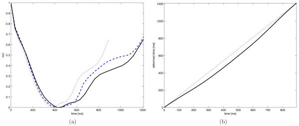
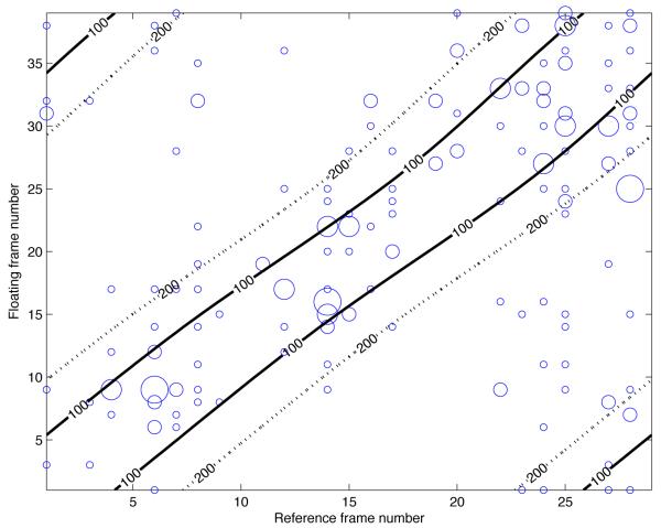
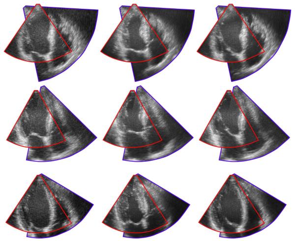
Registration of multiple 3D ultrasound sectors in order to provide an extended field of view is important for the appreciation of larger anatomical structures at high spatial and temporal resolution. In this paper, we present a method for fully automatic spatio-temporal registration between two partially overlapping 3D ultrasound sequences. The temporal alignment is solved by aligning the normalized cross correlation-over-time curves of the sequences. For the spatial alignment, corresponding 3D Scale Invariant Feature Transform (SIFT) features are extracted from all frames of both sequences independently of the temporal alignment. A rigid transform is then calculated by least squares minimization in combination with random sample consensus. The method is applied to 16 echocardiographic sequences of the left and right ventricles and evaluated against manually annotated temporal events and spatial anatomical landmarks. The mean distances between manually identified landmarks in the left and right ventricles after automatic registration were (mean ± SD) 4.3 ± 1.2 mm compared to a reference error of 2.8 ± 0.6 mm with manual registration. For the temporal alignment, the absolute errors in valvular event times were 14.4 ± 11.6 ms for Aortic Valve (AV) opening, 18.6 ± 16.0 ms for AV closing, and 34.6 ± 26.4 ms for mitral valve opening, compared to a mean inter-frame time of 29 ms.
Figures
Similar articles
-
[Study on the method of automatically determining maxillary complex landmarks based on non-rigid registration algorithms].Zhonghua Kou Qiang Yi Xue Za Zhi. 2023 Jun 9;58(6):554-560. doi: 10.3760/cma.j.cn112144-20230218-00053. Zhonghua Kou Qiang Yi Xue Za Zhi. 2023. PMID: 37272000 Chinese.
-
Quantification of organ motion based on an adaptive image-based scale invariant feature method.Med Phys. 2013 Nov;40(11):111701. doi: 10.1118/1.4822486. Med Phys. 2013. PMID: 24320409
-
Spatio-temporal (2D+T) non-rigid registration of real-time 3D echocardiography and cardiovascular MR image sequences.Phys Med Biol. 2011 Mar 7;56(5):1341-60. doi: 10.1088/0031-9155/56/5/008. Epub 2011 Feb 4. Phys Med Biol. 2011. PMID: 21297241
-
Non-rigid image registration using a modified fuzzy feature-based inference system for 3D cardiac motion estimation.Comput Methods Programs Biomed. 2021 Jun;205:106085. doi: 10.1016/j.cmpb.2021.106085. Epub 2021 Apr 6. Comput Methods Programs Biomed. 2021. PMID: 33878531
-
Spatiotemporal registration of multiple three-dimensional echocardiographic recordings for enhanced field of view imaging.J Med Imaging (Bellingham). 2016 Jul;3(3):037001. doi: 10.1117/1.JMI.3.3.037001. Epub 2016 Jul 8. J Med Imaging (Bellingham). 2016. PMID: 27446972 Free PMC article.
Cited by
-
Non-rigid registration of 3D ultrasound for neurosurgery using automatic feature detection and matching.Int J Comput Assist Radiol Surg. 2018 Oct;13(10):1525-1538. doi: 10.1007/s11548-018-1786-7. Epub 2018 Jun 4. Int J Comput Assist Radiol Surg. 2018. PMID: 29869321 Free PMC article.
-
Uncertainty-aware asynchronous scattered motion interpolation using Gaussian process regression.Comput Med Imaging Graph. 2019 Mar;72:1-12. doi: 10.1016/j.compmedimag.2018.12.001. Epub 2018 Dec 21. Comput Med Imaging Graph. 2019. PMID: 30654093 Free PMC article.
-
Semiautomated biventricular segmentation in three-dimensional echocardiography by coupled deformable surfaces.J Med Imaging (Bellingham). 2017 Apr;4(2):024005. doi: 10.1117/1.JMI.4.2.024005. Epub 2017 May 24. J Med Imaging (Bellingham). 2017. PMID: 28560243 Free PMC article.
-
Deformable multimodal registration for navigation in beating-heart cardiac surgery.Int J Comput Assist Radiol Surg. 2019 Jun;14(6):955-966. doi: 10.1007/s11548-019-01932-2. Epub 2019 Mar 19. Int J Comput Assist Radiol Surg. 2019. PMID: 30888597
References
-
- Ledesma-Carbayo M, Kybic J, Desco M, Santos A, Suhling M, Hunziker P, Unser M. Spatio-temporal nonrigid registration for ultrasound cardiac motion estimation. Medical Imaging, IEEE Transactions on. 2005 Sep;24:1113–1126. - PubMed
-
- Elen A, Choi HF, Loeckx D, Gao H, Claus P, Suetens P, Maes F, D’hooge J. Three-dimensional cardiac strain estimation using spatio-temporal elastic registration of ultrasound images: A feasibility study. Medical Imaging, IEEE Transactions on. 2008 Nov;27:1580–1591. - PubMed
-
- Kiss G, Barbosa D, Hristova K, Crosby J, Orderud F, Claus P, Amundsen B, Loeckx D, D’hooge J, Torp H. [Ultrasonics Symposium (IUS), 2009 IEEE International] Sep, 2009. Assessment of regional myocardial function using 3d cardiac strain estimation: comparison against conventional echocardiographic assessment; pp. 507–510.
-
- De Craene M, Piella G, Camara O, Duchateau N, Silva E, Doltra A, D’hooge J, Brugada J, Sitges M, Frangi A. Temporal diffeomorphic free-form deformation: application to motion and strain estimation from 3D echocardiography. Medical image analysis. 2012 Feb.16:427–50. - PubMed
-
- Danudibroto A, Gerard O, Alessandrini M, Mirea O, D’hooge J, Samset E. [Functional Imaging and Modeling of the Heart], Lecture Notes in Computer Science. Vol. 9126. Springer International Publishing; 2015. 3d farnebäck optic flow for extended field of view of echocardiography; pp. 129–136.
Grants and funding
LinkOut - more resources
Full Text Sources
Other Literature Sources