. 2015 Jul 22;25(28):4379-4389.
doi: 10.1002/adfm.201500875. Epub 2015 Jun 5.

Protein Corona Influences Cell-Biomaterial Interactions in Nanostructured Tissue Engineering Scaffolds

Affiliations

Protein Corona Influences Cell-Biomaterial Interactions in Nanostructured Tissue Engineering Scaffolds

Vahid Serpooshan et al. Adv Funct Mater. .

Abstract

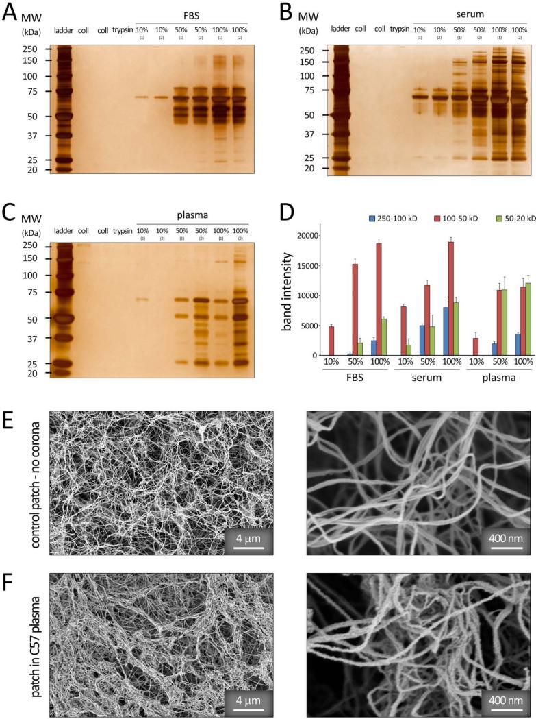
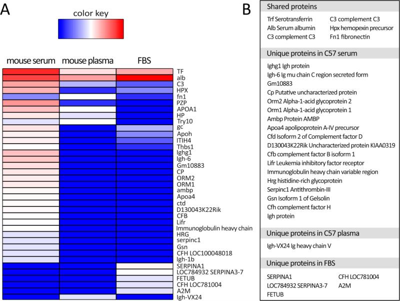
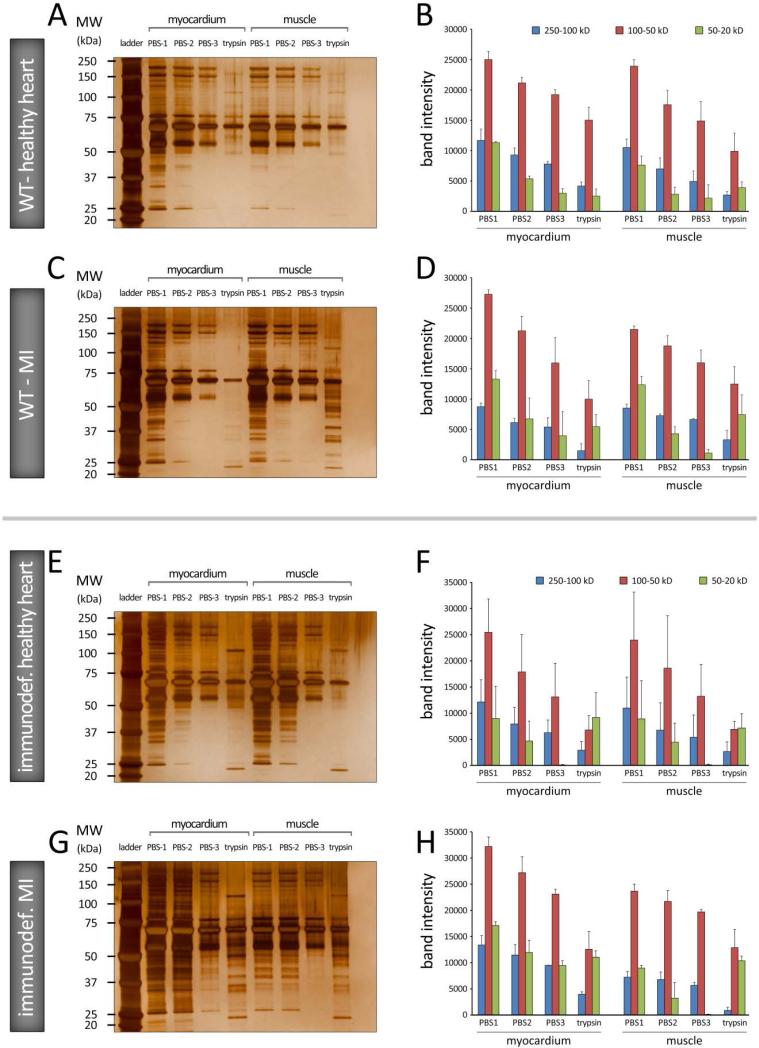
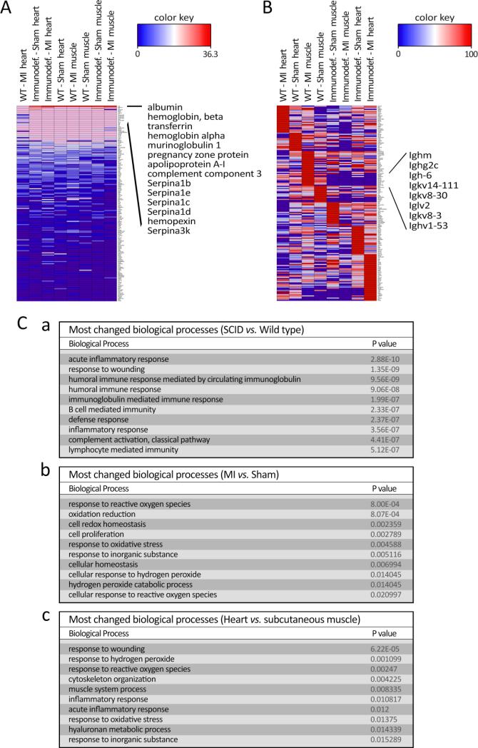
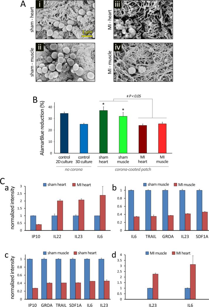
Biomaterials are extensively used to restore damaged tissues, in the forms of implants (e.g. tissue engineered scaffolds) or biomedical devices (e.g. pacemakers). Once in contact with the physiological environment, nanostructured biomaterials undergo modifications as a result of endogenous proteins binding to their surface. The formation of this macromolecular coating complex, known as 'protein corona', onto the surface of nanoparticles and its effect on cell-particle interactions are currently under intense investigation. In striking contrast, protein corona constructs within nanostructured porous tissue engineering scaffolds remain poorly characterized. As organismal systems are highly dynamic, it is conceivable that the formation of distinct protein corona on implanted scaffolds might itself modulate cell-extracellular matrix interactions. Here, we report that corona complexes formed onto the fibrils of engineered collagen scaffolds display specific, distinct, and reproducible compositions that are a signature of the tissue microenvironment as well as being indicative of the subject's health condition. Protein corona formed on collagen matrices modulated cellular secretome in a context-specific manner ex-vivo, demonstrating their role in regulating scaffold-cellular interactions. Together, these findings underscore the importance of custom-designing personalized nanostructured biomaterials, according to the biological milieu and disease state. We propose the use of protein corona as in situ biosensor of temporal and local biomarkers.

Keywords: collagen; in situ biosensor; personalized nanostructured biomaterials; protein corona; tissue engineering scaffold.


Figures

Figure 1
Figure 1
Schematic illustration of the patch-corona study; A: Plastic compression of highly hydrated collagen gels was used to generate a dense fibrillar scaffold structure (i.e. collagen patch).[38,44] The panel on the right shows scanning electron microscopy of the collagen patch fibrillar ultrastructure. B: Collagen scaffolds were incubated in different biological environments including i: fetal bovine serum, and C57 mouse serum and plasma, in vitro, ii: onto the sham (healthy) or myocardial infarcted (MI) heart tissue in C57 mouse, in vivo, and iii: onto the subcutaneous muscle tissue in healthy or MI C57 mouse, in vivo. C: two hours post incubation under each condition (i-iii), patches were removed and the protein coronas formed onto the scaffold were analyzed using SEM, SDS-PAGE, LC-MS/MS, and Luminex (human cytokines) immunoassay.
Figure 2
Figure 2
1D SDS–PAGE hard-corona protein profile for collagen patches incubated with various concentrations of (A) FBS, (B) C57 mouse serum, and (C) C57 mouse plasma proteins. The molecular weights (KDa) of the proteins in the standard ladder are reported on the left for reference. D: Histogram demonstrating the total band intensity of proteins recovered from various corona patches in (A-C). E-F: SEM images of the collagen patch before (E) and after incubation in vitro (in C57 plasma, F) confirming the adsorption of various protein macromolecules onto the collagen fibrils.
Figure 3
Figure 3
A: Heatmap data visualization of spectral counts of the associated proteins in the corona compositions of various in vitro conditions (C57 mouse serum, C57 muse plasma, and FBS), obtained by mass spectrometry. B: Table lists the proteins shared by C57 serum, plasma and FBS, and proteins unique to each condition.
Figure 4
Figure 4
A-D: 1D SDS–PAGE hard-corona protein profile for collagen patches incubated in vivo, in WT C57 BL/6J mice, under healthy heart (A and B) and myocardial infarction (MI) conditions (C and D) conditions. E-H: 1D SDS–PAGE hard-corona protein profile for patches incubated in immunodeficient C57 (SCID Beige) mice, under healthy heart (E and F) and MI (G and H) conditions. Each gel image shows the protein corona information of the patches grafted on myocardial (left) and subcutaneous muscle (right) tissues. The molecular weights (KDa) of the proteins in the standard ladder are reported on the left for reference. B, D, F, and H: Histograms demonstrating the total band intensity of proteins recovered from various corona patches in A, C, E, and G, respectively.
Figure 5
Figure 5
Heatmap data visualization of spectral counts of the associated proteins in corona composition obtained from different in vivo conditions: A: showing the total spec count ranking obtained by global normalization; and B: demonstrating expression ranking of normalized individual proteins which associated in corona composition of each in vivo condition. C: tables listing P values of top 10 significantly changed biological processes between (a) SCID vs. wild type, (b) MI vs. sham, and (c) heart vs. subcutaneous muscle, ranked by P value.
Figure 6
Figure 6
Evaluation of cells interaction with protein corona-patch following 24 hours of culture in vitro. Human umbilical vein endothelial cells (HUVECs) were plated onto collagen patches that were previously incubated in different in vivo conditions for 2 hours. HUVECs were also cultured on 2D substrate, and on 3D collagen patches, without protein corona, as controls. A: SEM images show HUVECs seeded onto the collagen patches incubated in i) Sham-heart, ii) Sham-muscle, iii) MI-heart, and iv) MI-muscle tissues. Remarkable differences were observed in the patch fibrillar ultrastructure, protein corona formation, and cell number/attachment. B: AlamarBlue assay demonstrated significantly different HUVEC viability/metabolism under varying conditions (P<0.05). *: significantly different compared to the 3D culture control (P<0.05). The differences in the means (± SEM) were tested using ANOVA to check for statistical significance (P < 0.05). C: the 63-plex Luminex Immunoassay of human cytokines detected significant differences in the production of multiple cytokines in the supernatant of HUVECs, after 24 hours of culture in vitro. Patch without cells was used to establish background signal in the assay; values smaller or not significantly greater than background were considered as negative. In all positively detected cytokines, those with levels significantly changed more than 2 folds between conditions are summarized in panels a-d.

References

    1. Monopoli MP, Åberg C, Salvati A, Dawson KA. Nat. Nanotechnol. 2012;7:779. - PubMed
    1. Monopoli MP, Bombelli FB, Dawson KA. Nat. Nanotechnol. 2011;6:11. - PubMed
    1. Lynch I, Salvati A, Dawson KA. Nat. Nanotechnol. 2009;4:546. - PubMed
    1. Laurent S, Burtea C, Thirifays C, Rezaee F, Mahmoudi M, Colloid Interface Sci J. 2013;392:431. - PubMed
    1. Mahmoudi M, Saeedi-Eslami SN, Shokrgozar MA, Azadmanesh K, Hassanlou M, Kalhor HR, Burtea C, Rothen-Rutishauser B, Laurent S, Sheibani S, Vali H. Nanoscale. 2012;4:5461. - PubMed

LinkOut - more resources