. 2016 Jul 28:10:356.
doi: 10.3389/fnins.2016.00356. eCollection 2016.

AAV Vectors for FRET-Based Analysis of Protein-Protein Interactions in Photoreceptor Outer Segments

Affiliations

AAV Vectors for FRET-Based Analysis of Protein-Protein Interactions in Photoreceptor Outer Segments

Elvir Becirovic et al. Front Neurosci. .

Abstract

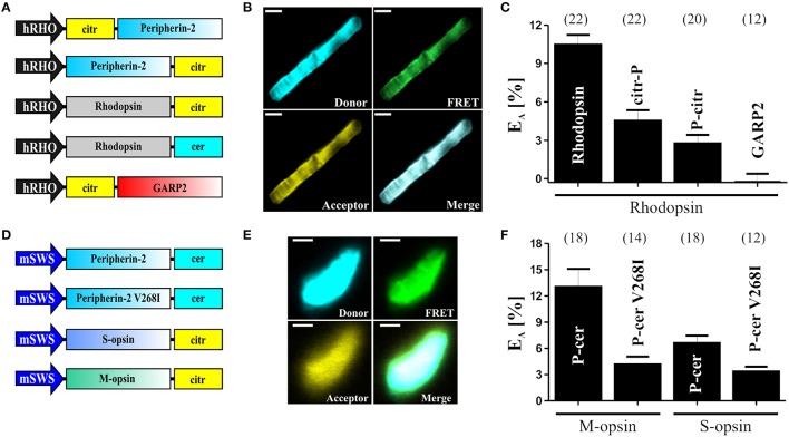
Fluorescence resonance energy transfer (FRET) is a powerful method for the detection and quantification of stationary and dynamic protein-protein interactions. Technical limitations have hampered systematic in vivo FRET experiments to study protein-protein interactions in their native environment. Here, we describe a rapid and robust protocol that combines adeno-associated virus (AAV) vector-mediated in vivo delivery of genetically encoded FRET partners with ex vivo FRET measurements. The method was established on acutely isolated outer segments of murine rod and cone photoreceptors and relies on the high co-transduction efficiency of retinal photoreceptors by co-delivered AAV vectors. The procedure can be used for the systematic analysis of protein-protein interactions of wild type or mutant outer segment proteins in their native environment. Conclusively, our protocol can help to characterize the physiological and pathophysiological relevance of photoreceptor specific proteins and, in principle, should also be transferable to other cell types.

Keywords: AAV; FRET; adeno-associated viral vectors; fluorescence resonance energy transfer; outer segment; photoreceptor; protein-protein interaction.


Figures

Figure 1
Figure 1
Schematic overview of the main steps of the procedure. AAV vectors encoding the FRET fusion proteins are produced in HEK293T cells (day 1–10). Single AAV vectors and combinations of AAVs encoding the FRET partners are delivered into the subretinal space of wildtype mice (day 11). Ten days later, OS are isolated for ex vivo FRET measurements.
Figure 2
Figure 2
OS preparation and FRET. (A) Overview of the protocol for the OS preparation and for the subsequent FRET measurements. (B) Representative images of isolated OS at different time points after the isolation. Scale bar, 3 μm.
Figure 3
Figure 3
FRET measurements in isolated photoreceptor OS. (A) Schematic depiction of the single constructs used for the subretinal injection and for the determination of the FRET efficiencies (EA) shown in (C). (B) Representative confocal images of single isolated rod OS co-expressing C-terminally tagged peripherin-2 and rhodopsin. The excitation wavelength and emission filter settings used to obtain the single channels (Donor, FRET and Acceptor) are summarized in the “Stepwise Procedures” section. Scale bar, 1.5 μm. (C) Results of the FRET measurements for different FRET pair combinations given as mean values ± standard error of the mean (SEM). Numbers of independent measurements (n) are given in brackets. EA values for the single FRET pairs are as follows: Rho-Rho, EA = 10.45 ± 0.78; Rho-citr-P, EA = 4.52 ± 0.82; Rho-P-citr, EA = 2.76 ± 0.65; Rho-GARP2, EA = 0.26 ± 0.65; (D) Schematic view of the single constructs used for the subretinal injection and for the determination of the FRET efficiencies (EA) shown in (F). (E) Representative confocal images of single isolated cone OS co-expressing C-terminally tagged peripherin-2 and cone opsin. Scale bar, 1.5 μm. (F) Results of the FRET measurements (given as mean values ± SEM) for different FRET pair combinations as indicated. Numbers of independent measurements (n) are given in brackets. EA values for the single FRET pairs are described elsewhere (Nguyen et al., 2016).

References

    1. Abe K., Zhao L., Periasamy A., Intes X., Barroso M. (2013). Non-invasive in vivo imaging of near infrared-labeled transferrin in breast cancer cells and tumors using fluorescence lifetime FRET. PLoS ONE 8:e80269. 10.1371/journal.pone.0080269 - DOI - PMC - PubMed
    1. Allocca M., Mussolino C., Garcia-Hoyos M., Sanges D., Iodice C., Petrillo M., et al. . (2007). Novel adeno-associated virus serotypes efficiently transduce murine photoreceptors. J. Virol. 81, 11372–11380. 10.1128/JVI.01327-07 - DOI - PMC - PubMed
    1. Arshavsky V. Y., Burns M. E. (2012). Photoreceptor signaling: supporting vision across a wide range of light intensities. J. Biol. Chem. 287, 1620–1626. 10.1074/jbc.R111.305243 - DOI - PMC - PubMed
    1. Auricchio A., Hildinger M., O'Connor E., Gao G. P., Wilson J. M. (2001). Isolation of highly infectious and pure adeno-associated virus type 2 vectors with a single-step gravity-flow column. Hum. Gene Ther. 12, 71–76. 10.1089/104303401450988 - DOI - PubMed
    1. Becirovic E., Bohm S., Nguyen O. N., Riedmayr L. M., Koch M. A., Schulze E., et al. . (2016). In vivo analysis of disease-associated point mutations unveils profound differences in mRNA splicing of peripherin-2 in rod and cone photoreceptors. PLoS Genet. 12:e1005811. 10.1371/journal.pgen.1005811 - DOI - PMC - PubMed