Traumatic brain injury increases levels of miR-21 in extracellular vesicles: implications for neuroinflammation
- PMID: 27516962
- PMCID: PMC4971839
- DOI: 10.1002/2211-5463.12092
Traumatic brain injury increases levels of miR-21 in extracellular vesicles: implications for neuroinflammation
Abstract
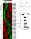
Traumatic brain injury (TBI) is an important health concern and effective treatment strategies remain elusive. Understanding the complex multicellular response to TBI may provide new avenues for intervention. In the context of TBI, cell-cell communication is critical. One relatively unexplored form of cell-cell communication in TBI is extracellular vesicles (EVs). These membrane-bound vesicles can carry many different types of cargo between cells. Recently, miRNA in EVs have been shown to mediate neuroinflammation and neuronal injury. To explore the role of EV-associated miRNA in TBI, we isolated EVs from the brain of injured mice and controls, purified RNA from brain EVs, and performed miRNA sequencing. We found that the expression of miR-212 decreased, while miR-21, miR-146, miR-7a, and miR-7b were significantly increased with injury, with miR-21 showing the largest change between conditions. The expression of miR-21 in the brain was primarily localized to neurons near the lesion site. Interestingly, adjacent to these miR-21-expressing neurons were activated microglia. The concurrent increase in miR-21 in EVs with the elevation of miR-21 in neurons, suggests that miR-21 is secreted from neurons as potential EV cargo. Thus, this study reveals a new potential mechanism of cell-cell communication not previously described in TBI.
Keywords: controlled cortical impact; exosomes; microRNA; microglia; neuroinflammation; secondary injury.
Figures


Similar articles
-
Extracellular Vesicles Derived From Neural Stem Cells, Astrocytes, and Microglia as Therapeutics for Easing TBI-Induced Brain Dysfunction.Stem Cells Transl Med. 2023 Mar 17;12(3):140-153. doi: 10.1093/stcltm/szad004. Stem Cells Transl Med. 2023. PMID: 36847078 Free PMC article. Review.
-
Extracellular Vesicles miRNA Cargo for Microglia Polarization in Traumatic Brain Injury.Biomolecules. 2020 Jun 12;10(6):901. doi: 10.3390/biom10060901. Biomolecules. 2020. PMID: 32545705 Free PMC article. Review.
-
Microglial-derived microparticles mediate neuroinflammation after traumatic brain injury.J Neuroinflammation. 2017 Mar 15;14(1):47. doi: 10.1186/s12974-017-0819-4. J Neuroinflammation. 2017. PMID: 28292310 Free PMC article.
-
Long noncoding RNA NKILA transferred by astrocyte-derived extracellular vesicles protects against neuronal injury by upregulating NLRX1 through binding to mir-195 in traumatic brain injury.Aging (Albany NY). 2021 Mar 3;13(6):8127-8145. doi: 10.18632/aging.202618. Epub 2021 Mar 3. Aging (Albany NY). 2021. PMID: 33686956 Free PMC article.
-
The Role of Microglial Exosomes and miR-124-3p in Neuroinflammation and Neuronal Repair after Traumatic Brain Injury.Life (Basel). 2023 Sep 16;13(9):1924. doi: 10.3390/life13091924. Life (Basel). 2023. PMID: 37763327 Free PMC article. Review.
Cited by
-
Extracellular vesicles in the diagnosis and treatment of central nervous system diseases.Neural Regen Res. 2020 Apr;15(4):586-596. doi: 10.4103/1673-5374.266908. Neural Regen Res. 2020. PMID: 31638080 Free PMC article. Review.
-
MicroRNA-21 Is a Versatile Regulator and Potential Treatment Target in Central Nervous System Disorders.Front Mol Neurosci. 2022 Jan 31;15:842288. doi: 10.3389/fnmol.2022.842288. eCollection 2022. Front Mol Neurosci. 2022. PMID: 35173580 Free PMC article. Review.
-
Human Lung Cell Pyroptosis Following Traumatic Brain Injury.Cells. 2019 Jan 18;8(1):69. doi: 10.3390/cells8010069. Cells. 2019. PMID: 30669285 Free PMC article.
-
Extracellular Vesicles: Packages Sent With Complement.Front Immunol. 2018 Apr 11;9:721. doi: 10.3389/fimmu.2018.00721. eCollection 2018. Front Immunol. 2018. PMID: 29696020 Free PMC article. Review.
-
Neutral Sphingomyelinase Inhibition Alleviates LPS-Induced Microglia Activation and Neuroinflammation after Experimental Traumatic Brain Injury.J Pharmacol Exp Ther. 2019 Mar;368(3):338-352. doi: 10.1124/jpet.118.253955. Epub 2018 Dec 18. J Pharmacol Exp Ther. 2019. PMID: 30563941 Free PMC article.
References
-
- Zaloshnja E, Miller T, Langlois JA and Selassie AW (2008) Prevalence of long‐term disability from traumatic brain injury in the civilian population of the United States, 2005. J Head Trauma Rehabil 23, 394–400. - PubMed
-
- Kuo CY, Liou TH, Chang KH, Chi WC, Escorpizo R, Yen CF, Liao HF, Chiou HY, Chiu WT and Tsai JT (2015) Functioning and disability analysis of patients with traumatic brain injury and spinal cord injury by using the world health organization disability assessment schedule 2.0. Int J Environ Res Public Health 12, 4116–4127. - PMC - PubMed
-
- Finnie JW (2013) Neuroinflammation: beneficial and detrimental effects after traumatic brain injury. Inflammopharmacology 21, 309–320. - PubMed
Grants and funding
LinkOut - more resources
Full Text Sources
Other Literature Sources

