. 2016 Aug 1;3(4):ENEURO.0150-16.2016.
doi: 10.1523/ENEURO.0150-16.2016. eCollection 2016 Jul-Aug.

APP Overexpression Causes Aβ-Independent Neuronal Death through Intrinsic Apoptosis Pathway

Affiliations

APP Overexpression Causes Aβ-Independent Neuronal Death through Intrinsic Apoptosis Pathway

Ning Cheng et al. eNeuro. .

Abstract

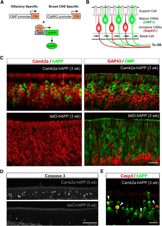
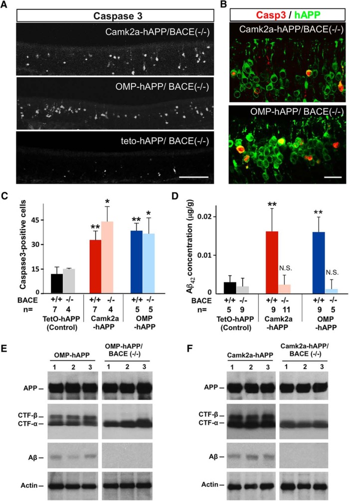
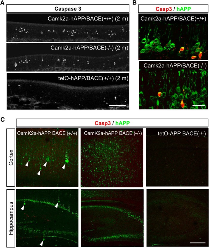
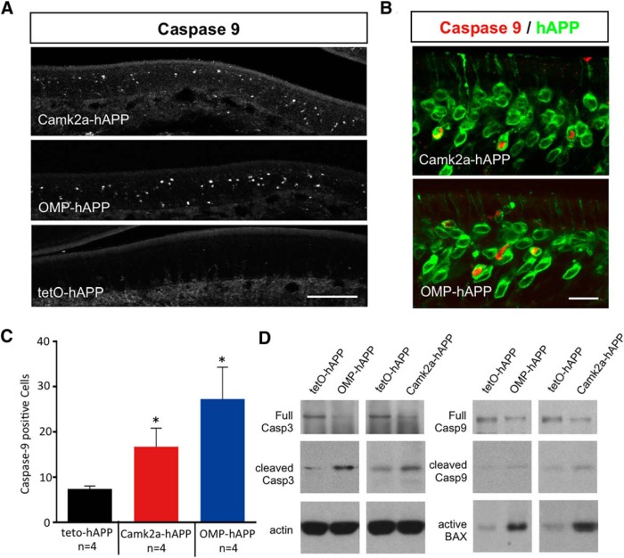
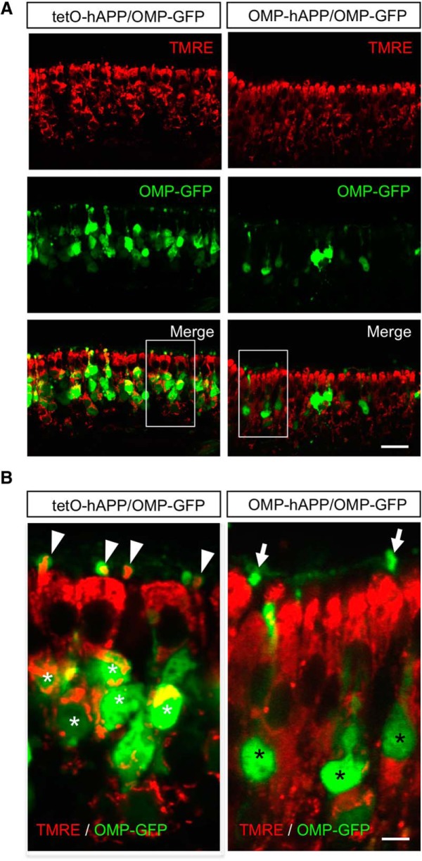
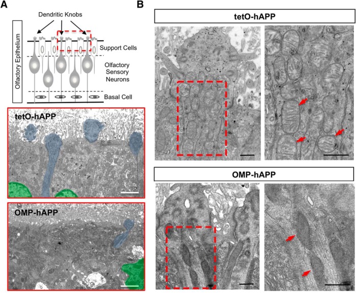
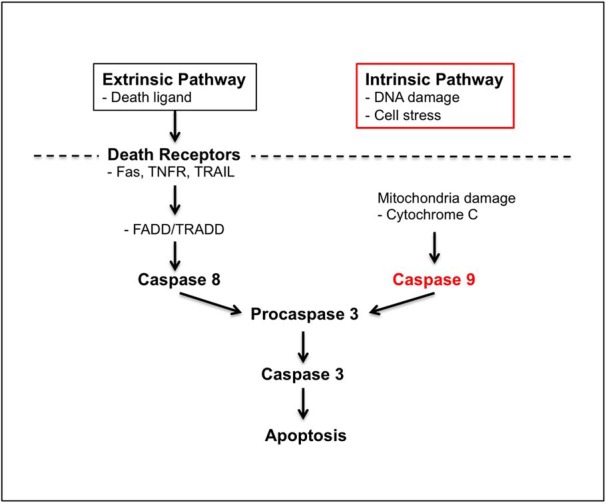
Accumulation of amyloid-β (Aβ) peptide in the brain is a central hallmark of Alzheimer's disease (AD) and is thought to be the cause of the observed neurodegeneration. Many animal models have been generated that overproduce Aβ yet do not exhibit clear neuronal loss, questioning this Aβ hypothesis. We previously developed an in vivo mouse model that expresses a humanized amyloid precursor protein (hAPP) in olfactory sensory neurons (OSNs) showing robust apoptosis and olfactory dysfunction by 3 weeks of age, which is consistent with early OSN loss and smell deficits, as observed in AD patients. Here we show, by deleting the β-site APP cleaving enzyme 1 (BACE1) in two distinct transgenic mouse models, that hAPP-induced apoptosis of OSNs is Aβ independent and remains cell autonomous. In addition, we reveal that the intrinsic apoptosis pathway is responsible for hAPP-induced OSN death, as marked by mitochondrial damage and caspase-9 activation. Given that hAPP expression causes OSN apoptosis despite the absence of BACE1, we propose that Aβ is not the sole cause of hAPP-induced neurodegeneration and that the early loss of olfactory function in AD may be based on a cell-autonomous mechanism, which could mark an early phase of AD, prior to Aβ accumulation. Thus, the olfactory system could serve as an important new platform to study the development of AD, providing unique insight for both early diagnosis and intervention.

Keywords: amyloid precursor protein; apoptosis; neurodegeneration; olfactory.


Figures

Figure 1.
Figure 1.
OSN apoptosis in Camk2a-hAPP mice. A, Strategies to generate mutant lines with olfactory-specific or broad CNS-specific overexpression of hAPP by using OMP or Camk2a promoter, respectively, and the tTA-TetO system. B, Diagram showing the organization of the OE, with markers for mature and immature OSNs highlighted. C, Left panels, Camk2a (red) and hAPP (green) immunohistochemical signal in the olfactory epithelium from 3-week-old Camk2a-hAPP and tetO-hAPP mice, respectively. Camk2a was broadly expressed in OSNs and mostly colocalized with hAPP in Camk2a-hAPP mice. Right panels, GAP43 (immature OSN marker, red) and OMP (mature OSN marker, green) immunohistochemical signal in the epithelium. Note that Camk2a-hAPP animals had less mature OSNs and thinner epithelia than controls. D, E, The 3-week-old Camk2a-hAPP animal had many more cleaved caspase-3-positive cells in the epithelium than the control animal (D), which colocalized in hAPP-expressing neurons (E, arrowheads). Scale bars: C, E, 20 µm; D, 100 µm.
Figure 2.
Figure 2.
hAPP-induced apoptosis persisted in a BACE−/− background. A, Cleaved caspase-3 signal in 3-week-old Camk2a-hAPP/BACE−/−, OMP-hAPP/BACE−/−, and control animals. B, The mutants had many more OSNs with caspase-3 signal than controls, which colocalized with the hAPP signal. C, Quantification of caspase-3-positive cells showed that hAPP-expressing lines had significantly more dying cells than the control lines, regardless of BACE genotype (Camk2a-hAPP, 32.8 ± 5.5, n = 7; and OMP-hAPP, 38.5 ± 4.6, n = 5, compared with tetO-hAPP, 11.7 ± 4.3, n = 7; while Camk2a-hAPP/BACE−/−, 44.1 ± 9.2, n = 4, and OMP-hAPP/BACE−/−, 36.3 ± 9.7, n = 5, compared with tetO-hAPP/BACE−/−, 14.8 ± 0.2, n = 4). D, ELISA on OE tissue shows very low levels of Aβ42 concentration in hAPP-expressing mutants with a null BACE background, not significantly different from the levels found in the control lines. E, F, Western blots confirming similar levels of full-length hAPP protein expression in the OEs from OMP-hAPP mice and OMP-hAPP/BACE−/− mice (E), as well as Camk2a-hAPP mice and Camk2a-hAPP/BACE−/− mice (F; individual animals in each lane). Actin was used as a loading control. The expression of α-CTF was also detected across all mutant genotypes, while β-CTF was absent in both the OMP-hAPP/BACE−/− mice and Camk2a-hAPP/BACE−/− mice, consistent with a loss in BACE activity. In addition, Aβ peptide is found in OEs from OMP-hAPP and Camk2a-hAPP mice, but is not detectable in OMP-hAPP/BACE−/− and Camk2a-hAPP/BACE−/− mice, further confirming the lack of BACE activity. All values are reported as the mean ± SD. Scale bars: A, 100 µm; B, 20 µm. *p < 0.01, **p < 0.001.
Figure 3.
Figure 3.
Cell death in the olfactory epithelium continued into adulthood without Aβ deposits. A, Cleaved caspase-3 signal in the OE of 2-month-old Camk2a-hAPP/BACE−/−, OMP-hAPP/BACE−/−, and control animals. B, At this age, the mutants still had many more OSNs with caspase-3 signal than controls, which colocalized with hAPP signal. C, Amyloid deposits developed in the cortex and hippocampus of 2-month-old Camk2a-hAPP/BACE+/+ mice (arrowheads), but not Camk2a-hAPP/BACE−/− or tetO-hAPP/BACE−/− mice. Cleaved caspase-3 signal was not elevated in the cortex or hippocampus of any genotype at either 3 weeks of age (data not shown) or 2 months of age. Scale bars: A, 100 µm; B, 20 µm; C, 200 µm.
Figure 4.
Figure 4.
Active caspase-9 expression in hAPP-expressing OSNs of both OMP-hAPP and Camk2a-hAPP mice. A, B, Cleaved caspase-9 signal in the OE of 3-week-old OMP-hAPP, Camk2a-hAPP, and control animals (A) showed OSNs that were positive for caspase-9 signal and clearly colocalized with hAPP signal shown in both mutant lines (B). C, Quantification of caspase-9-positive cells showed a significant increase in both hAPP-expressing lines compared with the controls (Camk2a-hAPP, 16.7 ± 4.1, n = 4; and OMP-hAPP, 27.3 ± 7.0, n = 4; compared with tetO-hAPP, 7.4 ± 0.6, n = 4). D, In addition, relative expression levels of the apoptosis markers, active caspase-3, caspase-9, and BAX were all increased in the mutant lines compared with controls, while full-length caspase-3 and caspase-9 levels showed a small reduction in mutant lines compared with control, which is consistent with an increased conversion to their cleaved active forms. Scale bars: A, 100 µm; B, 20 µm. *p < 0.05.
Figure 5.
Figure 5.
Vital-dye staining indicates dysfunctional mitochondria in hAPP-expressing OSNs. A, Fluorescent images of olfactory epithelium from OMP-hAPP mutant mice (right panels) and tetO-hAPP controls (left panels) comparing in vivo mitochondrial staining via TMRE indicator (red) in OMP-GFP-positive OSNs (green). B, Close-up of boxed regions in A showing that OSNs from control mice (left) contain live mitochondria (white asterisks) that are also detectable in the OSN dendritic knobs (arrowhead). By comparison, OSNs from mutant mice (right) show little colocalization with TMRE signal both in cell bodies (black asterisks) and in dendritic knobs (arrows). Scale bars: A, 20 µm; B, 5 µm.
Figure 6.
Figure 6.
Ultrastructural imaging of OE shows damaged mitochondria in dendritic knobs of hAPP-expressing OSNs. A, Schematic of OE depicting the superficial sustentacular support cells lining the apical surface with OSN dendritic knobs protruding between them into the lumen. Electron micrographs corresponding to the OE apical region (red boxed region in schematic) with tetO-hAPP control (middle) showing three support cells, two with distinct nuclei (shaded green), and portions of four OSN dendritic knobs (shaded blue) protruding into the lumen among the fragmented cilia, while an OMP-hAPP mutant (bottom) shows disrupted support cell organization (green nucleus) and very few OSN dendritic knobs (one shaded blue). B, Comparison of an OSN dendritic knob from tetO-hAPP control (top panels) and OMP-hAPP mutant (bottom panels) reveals a clear alteration in the mitochondrial morphology of OMP-hAPP animals, which appear dark with indistinct features compared with the healthy appearance of mitochondria in the control animals showing clear cisternae. The panels on the right correspond to the boxed regions on the left panels. Arrowheads point to mitochondria. Scale bars: A, 2 µm; B, 500 nm.
Figure 7.
Figure 7.
hAPP expression activates the intrinsic apoptosis pathway in OSNs. Schematic showing the key steps in both extrinsic and intrinsic pathways of apoptosis. Both intrinsic and extrinsic apoptosis pathways can lead to cell death by activating common late stage factors, such as caspase-3 but may be triggered through distinct mechanisms. The extrinsic pathway is typically activated via death receptors in the plasma membrane [e.g., Fas, TNFR (tumor necrosis factor receptor), TRAIL (tumor necrosis factor-related apoptosis-inducing ligand)], which activate FADD (FAS-associated death domain)/TRADD (Tumor necrosis factor receptor type 1-associated death domain), leading to caspase-8-mediated activation of the late-stage apoptosis pathway. The intrinsic pathway can be triggered by cell-autonomous factors (e.g., DNA damage, cell stress) possibly involving mitochondrial dysfunction and activation of caspase-9, which can activate caspase-3 followed by apoptosis. The clear activation of caspase-9 in hAPP-expressing OSNs both in OMP-hAPP and Camk2a-hAPP mutant mouse lines combined with the overt mitochondrial changes strongly indicate that hAPP-induced apoptosis is mediated by the intrinsic pathway.

Similar articles

Cited by

References

    1. Alzheimer's Association (2013) 2013 Alzheimer's disease facts and figures. Alzheimers Dement 9:208–245. 10.1016/j.jalz.2013.02.003 - DOI - PubMed
    1. Arnold SE, Lee EB, Moberg PJ, Stutzbach L, Kazi H, Han LY, Lee VM, Trojanowski JQ (2010) Olfactory epithelium amyloid-beta and paired helical filament-tau pathology in Alzheimer disease. Ann Neurol 67:462–469. 10.1002/ana.21910 - DOI - PMC - PubMed
    1. Bacon AW, Bondi MW, Salmon DP, Murphy C (1998) Very early changes in olfactory functioning due to Alzheimer's disease and the role of apolipoprotein E in olfaction. Ann N Y Acad Sci 855:723–731. - PubMed
    1. Behl C (2000) Apoptosis and Alzheimer's disease. J Neural Transm (Vienna) 107:1325–1344. 10.1007/s007020070021 - DOI - PubMed
    1. Cai H, Wang Y, McCarthy D, Wen H, Borchelt DR, Price DL, Wong PC (2001) BACE1 is the major beta-secretase for generation of Abeta peptides by neurons. Nat Neurosci 4:233–234. 10.1038/85064 - DOI - PubMed

Publication types

MeSH terms

LinkOut - more resources