Review
. 2016 Oct:33:131-139.
doi: 10.1016/j.mib.2016.06.007. Epub 2016 Aug 10.

An intracellular compass spatially coordinates cell cycle modules in Caulobacter crescentus

Affiliations
Review

An intracellular compass spatially coordinates cell cycle modules in Caulobacter crescentus

Keren Lasker et al. Curr Opin Microbiol. 2016 Oct.

Abstract

Cellular functions in Bacteria, such as chromosome segregation and cytokinesis, result from cascades of molecular events operating largely as self-contained modules. Regulated timing of these cellular modules stems from global genetic circuits that allow precise temporal activation with respect to cell cycle progression and cell differentiation. Critically, many of these functions occur at defined locations within the cell, and therefore regulators of each module must communicate to remain coordinated in space. In this perspective, we highlight recent discoveries in Caulobacter crescentus asymmetric cell division to illuminate diverse mechanisms by which a cellular compass, composed of scaffolding and signaling proteins, directs cell cycle modules to their exact cellular addresses.


Conflict of interest statement

The authors declare no competing interests.

Figures

Figure 1
Figure 1
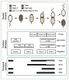
C. crescentus cell cycle control architecture. (a) The C. crescentus cell cycle can be viewed as a set of cellular modules (blue box), coordinated spatially (brown box) and temporally (green box). Top panel, Spatial Control. The swarmer cell has a single polar flagellum. The phosphatase PleC (orange) is localized at the flagellar pole. The swarmer cell begins to differentiate into a stalked cell. PleC becomes diffuse and is replaced at the differentiating pole with the kinase DivJ (blue). Activation of DivJ leads to the initiation of DNA replication. During this differentiation period, ejection of the flagellum permits construction of a stalk at the same cell pole. Chromosome replication and segregation then proceed simultaneously as the cell grows into a predivisional state. During this time, the cell begins assembling the cytokinesis machinery, whose core is the polymerizing GTPase FtsZ, at midcell (green). At the new cell pole, the de novo construction of a single flagellum occurs concurrently with the assembly of a set of scaffolding and signaling proteins, including PleC. As chromosome replication completes, the FtsZ ‘Z-ring’ constricts and disassembles, closing the inner membrane and separating the cytoplasm into two compartments. Cytoplasmic compartmentalization sequesters multiple signaling factors, including PleC at the new pole and DivJ at the old pole, enabling each chamber to initiate divergent genetic programs before full separation of the daughter cells. The cell type-specific presence of the master regulator CtrA, a downstream target of DivJ and PleC activity, is shown in grayscale to represent its abundance over the course of the cell cycle. Middle panel, Cellular Modules. Specific cell cycle events are shown as ‘Cellular Modules.’ Seven cellular modules are highlighted here; many more exist and have been omitted for clarity. Bottom panel, Temporal Control. Black bars indicate the abundance time period across the cell cycle for the master regulators DnaA, CcrM, GcrA, CtrA, SciP and MucR1/MucR2. Following cell division, the black top half of the bar reflects levels in the swarmer progeny and the black lower half of the bar reflects levels in the stalked progeny. Note that all master regulators except for MucR1/MucR2 are under cell cycle transcriptional control. (b) A temporally controlled transcription circuit (green) interfaces with spatially resolved signaling mechanisms (brown) to coordinate modular cellular processes (blue). Arrows indicate the connection between the modules and the proteins at the interface. Five cellular modules are highlighted here; many more exist and have been omitted for clarity. The genetic circuit guides the timing of expression of genes in all five modules. The activation and inhibition relationships between DnaA, CcrM, GcrA, CtrA, SciP, and MucR1/2 are illustrated. Spatial control results from the differential localization of distinct scaffolding proteins, which localize signaling proteins to the cell poles to generate cellular compartment-specific signaling states. Members of the compartment-sensing component (single domain response regulator DivK and diguanylate cyclase PleD) are modulated by two membrane histidine kinases PleC and DivJ, that act as swarmer and stalked cell determinants, respectively, each residing in a different pole. The compartment-sensing component modulates the localization of the flagellum and the stalk and also provides inputs for the asymmetry determination module. Proteins involved in the asymmetry determination module are shown in Figure 2a.
Figure 2
Figure 2
Regulation of CtrA levels and activity. (a) A more detailed pathway diagram of the regulatory network driving CtrA activation (orange) and CtrA degradation (blue), which was simplified in Figure 1a. Highlighted in brown: polar scaffolds PodJ and SpmX and the compartment-sensors PleD and DivK. The members of the pathways communicate via activation, inhibition, synthesis (of cdG), localization, transcriptional control (by CtrA and TacA), and degradation (by ClpXP and PdeA). Different arrowheads represent these distinct communication modes. (b) Snapshots of ClpXP dependent proteolysis as a function of cell cycle progression. Dark gray boxes represent active signaling function; white boxes represent a lack of activity. Divided boxes represent compartment-specific activity. Top: approximately 90 minutes into the cell cycle, ClpXP (purple and brown) degrades FtsZ (green) at the division plane. PleC phosphatase activity maintains low cdG levels and high CckA activity. High CckA activity maintains high CpdR~P levels, the inactive form. Middle: once compartmentalization completes, PleC is no longer present in the stalked compartment, deactivating CckA and promoting unphosphorylated CpdR (light brown) specifically in the stalked compartment. Unphosphorylated CpdR localizes ClpXP to the stalked pole and promotes degradation of the cdG phosphodiesterase, PdeA (dark blue). Bottom: once PdeA is degraded, cdG levels can rise again in the stalked compartment, allowing PopA to bind cdG and, with RcdA (light brown), direct CtrA (gray) to ClpXP for degradation.
Figure 3
Figure 3
Regulation of flagellum assembly and ejection. Top panel, Spatial Control. A diagram of the C. crescentus cell cycle highlights proteins that spatially regulate flagellum assembly and ejection. The distributions of FtsZ (green), cdG (brown), PleD (light blue), TipF (dark blue), and TipN (red) are shown throughout the cell cycle. The swarmer cell, with low cdG, has no TipF, as TipF is degraded when not bound to cdG. Upon differentiation and rising levels of cdG, TipF begins to accumulate with TipN at the new pole. TipF accumulation at the new pole permits recruitment of the initial flagellar base components. Z-ring assembly in early predivisional cells eventually leads to a relocalization of TipN and TipF at the division plane, defining the future ‘new poles’ for the incipient daughter cells; concurrently, the cascade leading to assembly of a new flagellum continues. Constriction of the Z-ring generates two separate cellular compartments. In the swarmer compartment, cdG levels decrease again, leading to degradation of TipF. FtsZ is also degraded as the cells prepare to separate. PleD does not localize to the cell poles when unphosphorylated and inactive. Middle panel, Cellular Modules. Events leading to flagellum assembly and ejection are highlighted. Lower panel, Temporal Control. Black bars indicate the abundance time period across the cell cycle for the flagellar regulators CtrA, GcrA, SciP and FlbD, with split white bars representing the lack of a factor in the nascent swarmer (top) or stalked (bottom) cell compartment.

References

    1. Kirkpatrick CL, Viollier PH. Decoding Caulobacter development. FEMS Microbiol Rev. 2012;36:193–205. - PubMed
    1. Laub MT, McAdams HH, Feldblyum T, Fraser CM, Shapiro L. Global analysis of the genetic network controlling a bacterial cell cycle. Science. 2000;290:2144–2148. - PubMed
    1. Zhou B, Schrader JM, Kalogeraki VS, Abeliuk E, Dinh CB, McAdams HH, Shapiro L. The global regulatory architecture of transcription during the caulobacter cell cycle. PLoS Genet. 2015;11:e1004831. Global analysis of Caulobacter transcription start sites reveals that many cell cycle controlled promoters respond to several of the master regulators. - PMC - PubMed
    1. Panis G, Murray SR, Viollier PH. Versatility of global transcriptional regulators in alpha-Proteobacteria: from essential cell cycle control to ancillary functions. FEMS Microbiol Rev. 2015;39:120–213. - PubMed
    1. Meier EL, Goley ED. Form and function of the bacterial cytokinetic ring. Curr Opin Cell Biol. 2014;26:19–27. - PubMed

Publication types

MeSH terms

LinkOut - more resources