. 2016 Sep 13;7(37):60230-60244.
doi: 10.18632/oncotarget.11176.

MicroRNA-126 inhibits colon cancer cell proliferation and invasion by targeting the chemokine (C-X-C motif) receptor 4 and Ras homolog gene family, member A, signaling pathway

Affiliations

MicroRNA-126 inhibits colon cancer cell proliferation and invasion by targeting the chemokine (C-X-C motif) receptor 4 and Ras homolog gene family, member A, signaling pathway

Wei Yuan et al. Oncotarget. .

Abstract

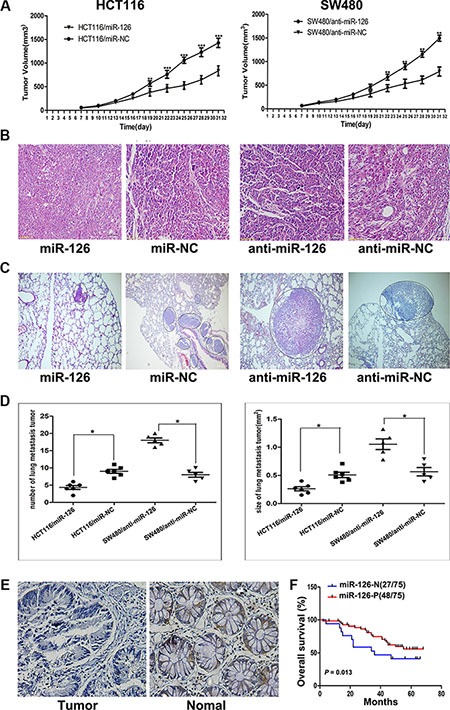
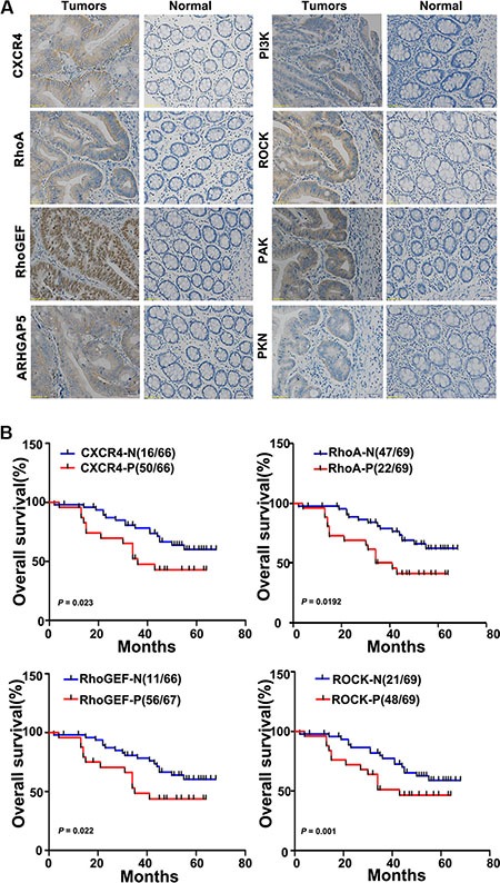
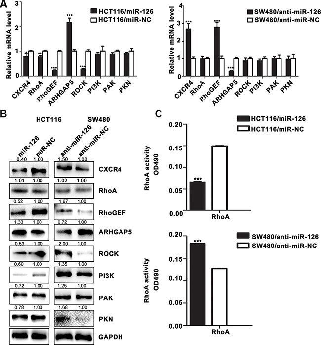
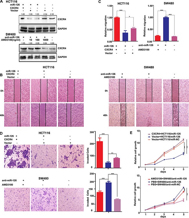
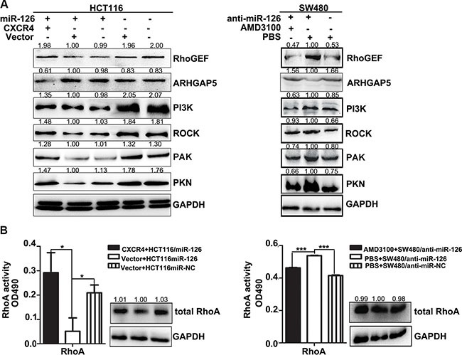
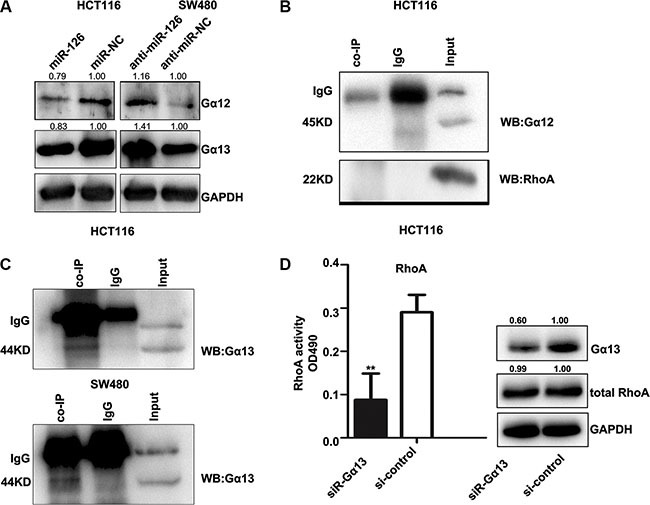
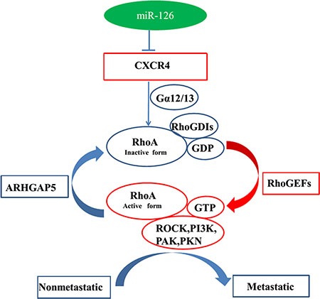
MicroRNA-126 (miR-126) suppresses the migration, proliferation and invasion of colon cancer cells. However, the underlying mechanisms of miR-126 in colon cancer have not been fully elucidated. In this study, in vivo experiments revealed that miR-126 inhibits colon cancer growth and metastasis. Furthermore, miR-126 was down-regulated in human colon cancer tissue, and its expression was inversely correlated with TNM stage and metastasis of patients. Low level of miR-126 identified patients with poor prognosis. And we found that miR-126 expression was negatively correlated with the expression levels of chemokine (C-X-C motif) receptor 4 (CXCR4) and components of signaling pathway of Ras homolog gene family, member A (RhoA) in vitro and in vivo. Moreover, we verified that miR-126 negatively regulated CXCR4 and RhoA signaling in vitro. In addition, either in miR-126-overexpressing or in miR- 126-silenced colon cancer cells, the restoration of CXCR4 could significantly reverse the proliferation and invasion, as well as abolish the effects of miR-126 on RhoA signaling pathway. Collectively, these results demonstrated that miR-126 acts as a tumor suppressor by inactivating RhoA signaling via CXCR4 in colon cancer. And miR-126 may serve as a prognostic marker for monitoring and treating colon cancer.

Keywords: CXCR4; RhoA; colon cancer; microRNA-126 (mir-126).


Conflict of interest statement

CONFLICTS OF INTEREST

The authors declare that they have no competing interests.

Figures

Figure 1
Figure 1. miR-126 inhibits colon cancer progression
(A) Tumor volume was measured 1 week after injection and every 3 d thereafter. The average tumor volume for HCT116/miR-126 was smaller than that of the control (HCT116/miR-NC; left panel; n = 5, p < 0.01, ANOVA), whereas the average tumor volume for SW480/anti-miR-126 was larger than that of the control (SW480/anti-miR-NC; right panel; n = 5, p < 0.01, ANOVA), **p < 0.01, ***p < 0.001. (B) Representative hematoxylin/eosin-stained images of subcutaneous tumors from mice that had been injected with transfected HCT116 or transfected SW480 cells (200×). (C) Representative hematoxylin/eosin-stained images of lung tissue sections from mice that had been injected with transfected HCT116 (left panel) or transfected SW480 cells (right panel). Black circles identify metastatic foci (200×). (D) Scatter diagrams depicting the average number (left panel) and size (right panel) of metastatic nodules in 4–5 microscope fields for each type of transfected cells, *p < 0.05. (E) A decrease in miR-126 was detected in microarrays of colon cancer tissues compared with that detected in normal colonic mucosa as shown by in situ hybridization (200×). (F) Kaplan-Meier survival curves for patients with different levels of miR-126 expression in their colon tumors. Red line: patients whose tissues expressed miR-126 (48/75 patients); blue line, patients whose tissues did not express miR-126 (27/75 patients). p = 0.013. MiR-126-N: miR-126 negative expression; miR-126-P: miR-126 positive expression.
Figure 2
Figure 2. CXCR4 and RhoA signaling pathway components are upregulated in colon cancer tissues and inversely correlate with the overall survival of colon cancer patients
(A) The relative levels of CXCR4 and the RhoA signaling pathway components RhoGEF, ARHGAP5, PI3K, ROCK, PAK, and PKN were assessed in colon cancer tissues and in paired normal colonic mucosa by immunohistochemical staining. Representative images are shown (200×). (B) Kaplan-Meier survival curves are shown for patients according to the expression of CXCR4, RhoA, RhoGEF, and ROCK in their colon cancer tissues. Colon cancer-associated death was significantly correlated with expression of CXCR4, RhoA, RhoGEF, and ROCK. Decreased patient overall survival was associated with higher expression of CXCR4 (upper left panel; n = 50/66; p = 0.023), RhoA (upper right panel; n = 47/69; p = 0.0192), RhoGEF (lower left panel; n = 56/67; p = 0.022), and ROCK (lower right panel; n = 48/69; p = 0.001). The red lines indicate patients with cancer tissues expressing CXCR4, RhoA, RhoGEF, and ROCK; blue lines indicate the patient tissues not expressing these proteins. In each graph, each ‘N’ implies negative expression, and each ‘P’ implies positive expression.
Figure 3
Figure 3. MiR-126 down-regulates the expression of CXCR4 and RhoA signaling pathway components in human colon cancer cells
(A) The mRNA levels of CXCR4 and components of the RhoA signaling pathway were determined by qRT-PCR in transfected HCT116 and SW480 cells. GAPDH expression served as the internal control.*p < 0.05; **p < 0.01; ***p < 0.001. (B) Western Blot for the detection of CXCR4 and components of the RhoA signaling pathway in transfected HCT116 and SW480 cells. GAPDH served as the internal control. (C) RhoA G-LISA activation assays were used to measure RhoA activity in the transfected cells. ***p < 0.001.
Figure 4
Figure 4. CXCR4 is a functional mediator for miR-126 in colon cancer cells
(A) Western Blots were used to confirm CXCR4 expression. Transient transfection of HCT116/miR-126 cells with CXCR4 plasmids resulted in increased expression of CXCR4 compared with that cells transfected with unmodified vectors or not transfected (upper panels). AMD3100 was used to inhibit CXCR4 expression in SW480/anti-miR-126 cells (bottom panels). At a concentration of 1000 ng/ml AMD3100, CXCR4 expression was substantially reduced. (B, C) Wound-healing assays were used to measure cell migration capacity. Migration increased following transient transfection with the CXCR4 plasmids (left panels), but the migration of SW480/anti-miR-126 cells was inhibited after exposure to AMD3100 (right panels) *p < 0.05, ***p < 0.001. (D) Transwell assays were used to measure cell invasion capacity. The invasion capacity of HCT116/miR-126 cells increased following transient transfection with the CXCR4 plasmids (top). The red, blue, and purple bars denote CXCR4 + HCT116/miR-126, vector + HCT116/miR-126, and vector + HCT116/miR-NC, respectively. The invasion capacity of SW480/anti-miR-126 cells was reduced after exposure to AMD3100 (bottom). The red, blue, and purple bars denote AMD3100 + SW480/anti-miR-126, PBS + SW480/anti-miR-126, and PBS + SW480/anti-miR-NC, respectively. Panels on the left show stained cells that had invaded. Cells in five randomly selected areas were counted, and a statistical analysis was performed using SPSS 17.0. (E) MTT assays were used to measure cell proliferation. The proliferation of HCT116/miR-126 cells was increased after transient transfection with the CXCR4 plasmids (top panel). The proliferation of SW480/anti-miR-126 cells was reduced after exposure to AMD3100 (bottom panel). The data represent the mean ± SD of three replicates **p < 0.01, ***p < 0.001.
Figure 5
Figure 5. CXCR4 abolishes the effects of miR-126 on RhoA signaling pathway in colon cancer
(A) Western blot analysis of the expression levels of components of the RhoA signaling pathway. Left panels show expression levels in HCT116/miR-126 cells after transient transfection with the CXCR4 plasmids. Right panels show expression levels in SW480/anti-miR-126 cells after exposure to AMD3100 (1000 ng/ml). (B) RhoA activation G-LISA assays were used to measure RhoA activity in HCT116/miR-126 cells transiently transfected with the CXCR4 plasmids (left panels) and in SW480/anti-miR-126 cells exposed to 1000 ng/ml AMD3100 (right panels). *p < 0.05, ***p < 0.001.
Figure 6
Figure 6. CXCR4 activates the RhoA signaling pathway via Gα13
(A) Western Blot showing the levels of Gα12 and Gα13 in transfected HCT116 and SW480 cells. GAPDH served as the internal control. (B, C) In vitro co-immunoprecipitation (co-IP) confirmed the interactions between CXCR4, RhoA, and the Gα12/Gα13 complex and revealed a complex composed of CXCR4 and Gα13. Total protein was extracted from untransfected HCT116 and SW480 cells. Total (input) and eluted (co-IP) proteins were subjected to Western Blot, which was performed with anti-Gα12, anti-RhoA, and anti-Gα13. (D) Western Blot was used to confirm the expression of Gα13 (right), and the G-LISA assay was used to measure RhoA activity in HCT116 cells after knocking down Gα13 expression with siRNA-Gα13 (left). The control group was exposed to the non-specific siRNA, **p < 0.01.
Figure 7
Figure 7. Model for miR-126-mediated inhibition of cell proliferation and migration via its regulation of the CXCR4 and RhoA signaling pathways
Rho GDP-dissociation inhibitors (RhoGDIs) are important regulators of the Rho family of small GTPases. RhoGDIs can prevent RhoA activation by inhibiting GDP dissociation from RhoA. RhoGEFs activate RhoA, and ARHGAP5 can negatively regulate RhoA. PAK, PKN, ROCK, and PI3K are downstream effectors of RhoA signaling. MiR-126 inhibits cell proliferation and migration via its regulation of the CXCR4/Gα13/RhoA signaling axis in colon cancer pathology.

Similar articles

Cited by

References

    1. Siegel R, Desantis C, Jemal A. Colorectal cancer statistics, 2014. CA Cancer J Clin. 2014;64:104–117. - PubMed
    1. Mulrane L, McGee SF, Gallagher WM, O'Connor DP. miRNA dysregulation in breast cancer. Cancer Res. 2013;73:6554–6562. - PubMed
    1. Bartel DP. MicroRNAs: genomics, biogenesis, mechanism, and function. Cell. 2004;116:281–297. - PubMed
    1. Calin GA, Croce CM. MicroRNA signatures in human cancers. Nat Rev Cancer. 2006;6:857–866. - PubMed
    1. Kumar MS, Lu J, Mercer KL, Golub TR, Jacks T. Impaired microRNA processing enhances cellular transformation and tumorigenesis. Nat Genet. 2007;39:673–677. - PubMed

MeSH terms

LinkOut - more resources