. 2016 Sep 13;7(37):60123-60132.
doi: 10.18632/oncotarget.11164.

SC79 protects retinal pigment epithelium cells from UV radiation via activating Akt-Nrf2 signaling

Affiliations

SC79 protects retinal pigment epithelium cells from UV radiation via activating Akt-Nrf2 signaling

Yi-Qing Gong et al. Oncotarget. .

Abstract

Excessive Ultra-violet (UV) radiation causes oxidative damages and apoptosis in retinal pigment epithelium (RPE) cells. Here we tested the potential activity of SC79, a novel small molecule activator of Akt, against the process. We showed that SC79 activated Akt in primary and established (ARPE-19 line) RPE cells. It protected RPE cells from UV damages possibly via inhibiting cell apoptosis. Akt inhibition, via an Akt specific inhibitor (MK-2206) or Akt1 shRNA silence, almost abolished SC79-induced RPE cytoprotection. Further studies showed that SC79 activated Akt-dependent NF-E2-related factor 2 (Nrf2) signaling and inhibited UV-induced oxidative stress in RPE cells. Reversely, Nrf2 shRNA knockdown or S40T mutation attenuated SC79-induced anti-UV activity. For the in vivo studies, we showed that intravitreal injection of SC79 significantly protected mouse retina from light damages. Based on these results, we suggest that SC79 protects RPE cells from UV damages possibly via activating Akt-Nrf2 signaling axis.

Keywords: Akt; SC79; UV; retinal pigment epithelium.


Conflict of interest statement

CONFLICTS OF INTEREST

None.

Figures

Figure 1
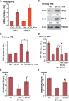
Figure 1. SC79 activates Akt and protects RPE cells from UV damages
The molecular structure along with the molecular weight (MW) of SC79 were presented (A). ARPE-19 cells were treated with applied concentration of SC79 (0.1–10 μg/mL, or 2.74–27.41 μM) for 1 h, p-Akt (Ser-473) and Akt1 expression was tested by Western blots and was quantified (B) n = 4). ARPE-19 cells (C and D) primary murine RPE cells (“Primary RPE”, (E)) or HLECs (F) pretreated with applied concentration of SC79 for 30 min, were subjected to UV radiation (30 mJ/cm2), cells were further cultured for 24 h, and cell viability was tested by MTT assay (C, E and F); Cell death was detected by trypan blue staining assay (D). “Ctrl” stands for untreated control group (Same for all figures). For each assay, n = 5. Experiments in this figure were repeated three times to insure consistency of results. *p < 0.05 vs. “Ctrl” group (C–F). **p < 0.05 vs. UV only group (C–F).
Figure 2
Figure 2. SC79 inhibits UV-induced apoptosis activation in RPE cells
ARPE-19 cells (AE) or primary murine RPE cells (“Primary RPE”,(F)) were pretreated with SC79 (5 μg/mL) for 30 min prior to UV radiation (30 mJ/cm2), cells were further cultured for applied time, and cell apoptosis was tested by listed assays (A–F). For each assay, n = 5. Experiments in this figure were repeated three times to insure consistency of results. *p < 0.05 vs. “Ctrl” group. **p < 0.05 vs. UV only group.
Figure 3
Figure 3. SC79-mediated RPE cytoprotection against UV requires Akt activation
ARPE-19 cells were pre-treated with MK-2206 (5 μM) for 1h, followed by SC79 (5 μg/mL) treatment for 1 h, p-Akt (Ser-473) and Akt1 expression was tested by Western blot assay (A). ARPE-19 cells were pre-treated with MK-2206 (5 μM) for 1 h, followed by UV radiation (30 mJ/cm2), or plus SC79 (5 μg/mL, 30 min prior UV), cells were further cultured for applied time; Cell viability ((B) MTT assay) and cell apoptosis ((C) Histone DNA ELISA assay) were tested. The stably ARPE-19 cells expressing scramble control shRNA (“Scr shRNA”) or Akt1 shRNA were treated with UV (30 mJ/cm2) radiation, or plus SC79 (5 μg/mL, 30 min prior UV); Cells were further cultured for applied time, cell viability (E) and cell apoptosis (F) were tested. SC79 (5 μg/mL, 1 h)-induced Akt activation in above cells was tested by Western blot assay (D). Akt phosphorylation (vs. regular Akt1) was quantified (A), Akt1 expression (vs. Tubulin) was quantified (D). “dmso” stands for 0.1% DMSO vehicle control (B and C). For each assay, n = 5. Experiments in this figure were repeated three times to insure consistency of results. *p < 0.05.
Figure 4
Figure 4. SC79 activates Nrf2 signaling in RPE cells
ARPE-19 cells were treated with applied concentration of SC79 (0.1–10 μg/mL) for 4 h, mRNA expression of HO-1, NQO-1 and Nrf2 was tested by RT-qPCR assay (A). Stably ARPE-19 cells with scramble-shRNA (“Scr shRNA”) or Akt1 shRNA were treated with SC79 (5 μg/mL) or plus MK-2206 (5 μM), HO-1 and NQO-1 mRNA expression was tested by RT-qPCR assay (B and C). ARPE-19 cells, pretreated with SC79 (5 μg/mL) for 30 min, were subjected to UV radiation (30 mJ/cm2), cells were further cultured and relative ROS production was tested (D). The stably ARPE-19 cells with scramble-shRNA (“Scr shRNA”) or Nrf2 shRNA (“−1/−2”, with non-overlapping sequences) were treated with SC79 (5 μg/mL) for indicated time, expressions of listed proteins and mRNA were tested by Western blot assay (E) and RT-qPCR assay (F and G) respectively. Above cells were treated with UV (30 mJ/cm2) radiation, or plus SC79 (5 μg/mL, 30 min prior UV), cells were further cultured for 24h and cell viability was evaluated by MTT assay (H). HO-1 protein expression (vs. Tubulin) was quantified (E). For each assay, n = 5. Experiments in this figure were repeated three times to insure consistency of results. *p < 0.05 vs. “Ctrl” group (A). *p < 0.05 (B, C, E and F).
Figure 5
Figure 5. Nrf2 S40T mutation attenuates SC79-mediated RPE cytoprotection against UV radiation
Stable ARPE-19 cells expressing dominant negative Nrf2 (S40T, “DN-Nrf2”, flag tagged) or empty vector (pSV2 puro Flag) were treated with SC79 (5 μg/mL) for indicated time, expressions of listed protein and mRNA were tested by Western blot assay (A) and RT-qPCR assay (B) respectively. Above cells were also subjected to UV (30 mJ/cm2) radiation, or plus SC79 (5 μg/mL, 30 min prior UV), cell viability and apoptosis were tested MTT assay (C) and Histone DNA ELISA assay (D) respectively. HO-1 protein expression (vs. Tubulin) was quantified (A). For each assay, n = 5. Experiments in this figure were repeated three times to insure consistency of results. *p < 0.05.
Figure 6
Figure 6. SC79 activates Nrf2 signaling in primary murine RPE cells and its retinal protection activity in vivo
Primary murine RPE cells were treated with SC79 (5 μg/mL) for indicated time, expressions of listed mRNAs (A) and proteins (B) were tested. Primary murine RPE cells were treated with UV (30 mJ/cm2) radiation, or plus SC79 (5 μg/mL, 30 min prior UV), cells were further cultured before ROS content was analyzed (C). Primary murine RPE cells were pre-treated with SC79 (5 μg/mL), or plus MK-2206 (“MK”, 5 μM)/ZnPP (10 μM), followed by UV (30 mJ/cm2) radiation, cells were further cultured for 24 h before cell viability was tested (D). After the light exposure in mice retina, ERG was measured, quantified amplitudes of a- and b-waves were presented (E and F). For each assay, n = 5. *p < 0.05 vs. “Ctrl” group (A, E and F). **p < 0.05 vs. “Light damage” only group (E and F). *p < 0.05 (C and D).

References

    1. van Lookeren Campagne M, LeCouter J, Yaspan BL, Ye W. Mechanisms of age-related macular degeneration and therapeutic opportunities. J Pathol. 2014;232:151–164. - PubMed
    1. Beatty S, Koh H, Phil M, Henson D, Boulton M. The role of oxidative stress in the pathogenesis of age-related macular degeneration. Surv Ophthalmol. 2000;45:115–134. - PubMed
    1. Young RW. Solar radiation and age-related macular degeneration. Surv Ophthalmol. 1988;32:252–269. - PubMed
    1. Cao G, Chen M, Song Q, Liu Y, Xie L, Han Y, Liu Z, Ji Y, Jiang Q. EGCG protects against UVB-induced apoptosis via oxidative stress and the JNK1/c-Jun pathway in ARPE19 cells. Mol Med Rep. 2012;5:54–59. - PubMed
    1. Yao J, Bi HE, Sheng Y, Cheng LB, Wendu RL, Wang CH, Cao GF, Jiang Q. Ultraviolet (UV) and hydrogen peroxide activate ceramide-ER stress-AMPK signaling axis to promote retinal pigment epithelium (RPE) cell apoptosis. Int J Mol Sci. 2013;14:10355–10368. - PMC - PubMed

MeSH terms

LinkOut - more resources