

Influence of Light Emitting Diode-Derived Blue Light Overexposure on Mouse Ocular Surface
- PMID: 27517861
- PMCID: PMC4982597
- DOI: 10.1371/journal.pone.0161041
Influence of Light Emitting Diode-Derived Blue Light Overexposure on Mouse Ocular Surface
Erratum in
-
Correction: Influence of Light Emitting Diode-Derived Blue Light Overexposure on Mouse Ocular Surface.PLoS One. 2016 Nov 30;11(11):e0167671. doi: 10.1371/journal.pone.0167671. eCollection 2016. PLoS One. 2016. PMID: 27902781 Free PMC article.
Abstract
Purpose: To investigate the influence of overexposure to light emitting diode (LED)-derived light with various wavelengths on mouse ocular surface.
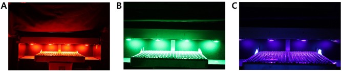
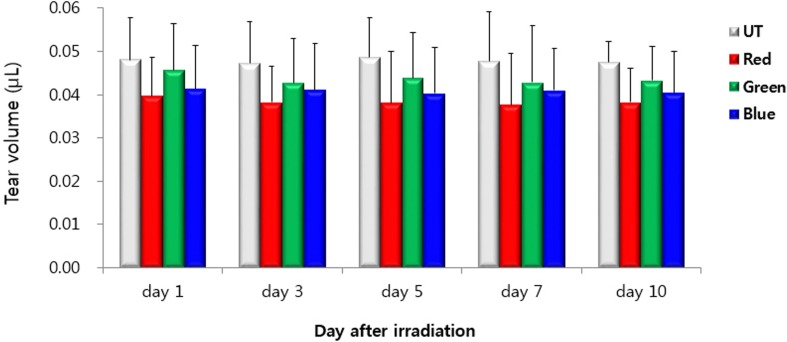
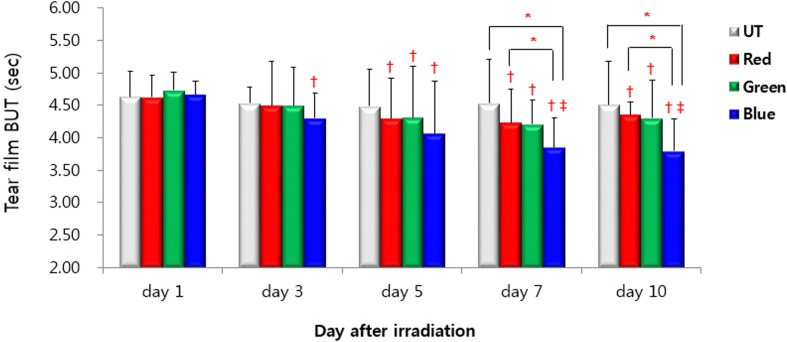
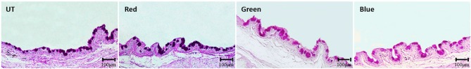
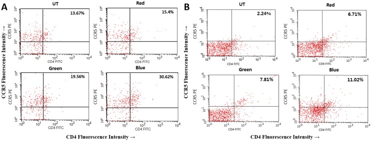
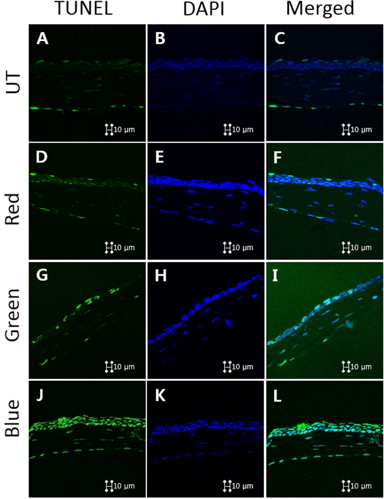
Methods: LEDs with various wavelengths were used to irradiate C57BL/6 mice at an energy dose of 50 J/cm2, twice a day, for 10 consecutive days. The red, green, and blue groups represented wavelengths of 630 nm, 525 nm, and 410 nm, respectively. The untouched group (UT) was not exposed to LED light and served as the untreated control. Tear volume, tear film break-up time (TBUT), and corneal fluorescein staining scores were measured on days 1, 3, 5, 7, and 10. Levels of interferon (IFN)-γ, interleukin (IL)-1β, IL-6, and tumor necrosis factor (TNF)-α were measured in the cornea and conjunctiva using a multiplex immunobead assay at day 10. Levels of malondialdehyde (MDA) were measured with an enzyme-linked immunosorbent assay. Flow cytometry, 2'7'-dichlorofluorescein diacetate (DCF-DA) assay, histologic analysis, immunohistochemistry with 4-hydroxynonenal, and terminal deoxynucleotidyl transferase-mediated dUTP-nick end labeling (TUNEL) staining were also performed.
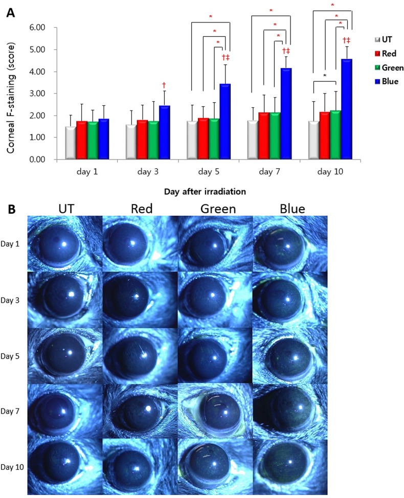
Results: TBUT of the blue group showed significant decreases at days 7 and 10, compared with the UT and red groups. Corneal fluorescein staining scores significantly increased in the blue group when compared with UT, red, and green groups at days 5, 7, and 10. A significant increase in the corneal levels of IL-1β and IL-6 was observed in the blue group, compared with the other groups. The blue group showed significantly increased reactive oxygen species production in the DCF-DA assay and increased inflammatory T cells in the flow cytometry. A significantly increased TUNEL positive cells was identified in the blue group.
Conclusions: Overexposure to blue light with short wavelengths can induce oxidative damage and apoptosis to the cornea, which may manifest as increased ocular surface inflammation and resultant dry eye.
Conflict of interest statement
Figures
Similar articles
-
Effects of eye drops containing a mixture of 3% diquafosol sodium and tocopherol acetate (vitamin E) on the ocular surface of murine dry eye.Cutan Ocul Toxicol. 2021 Dec;40(4):350-358. doi: 10.1080/15569527.2021.1973022. Epub 2021 Sep 8. Cutan Ocul Toxicol. 2021. PMID: 34496685
-
Effectiveness of topical infliximab in a mouse model of experimental dry eye.Cornea. 2012 Nov;31 Suppl 1:S25-31. doi: 10.1097/ICO.0b013e31826a80ea. Cornea. 2012. PMID: 23038030
-
Expression and Role of Nucleotide-Binding Oligomerization Domain 2 (NOD2) in the Ocular Surface of Murine Dry Eye.Invest Ophthalmol Vis Sci. 2019 Jun 3;60(7):2641-2649. doi: 10.1167/iovs.19-27144. Invest Ophthalmol Vis Sci. 2019. PMID: 31237655
-
Therapeutic application of light emitting diode: Photo-oncomic approach.J Photochem Photobiol B. 2019 Mar;192:1-7. doi: 10.1016/j.jphotobiol.2019.01.003. Epub 2019 Jan 9. J Photochem Photobiol B. 2019. PMID: 30654264 Review.
-
Mechanisms of blue light-induced eye hazard and protective measures: a review.Biomed Pharmacother. 2020 Oct;130:110577. doi: 10.1016/j.biopha.2020.110577. Epub 2020 Aug 4. Biomed Pharmacother. 2020. PMID: 32763817 Review.
Cited by
-
Whole transcriptome analysis on blue light-induced eye damage.Int J Ophthalmol. 2020 Aug 18;13(8):1210-1222. doi: 10.18240/ijo.2020.08.06. eCollection 2020. Int J Ophthalmol. 2020. PMID: 32821674 Free PMC article.
-
Diurnal variation of human tear meniscus volume measured with tear strip meniscometry self-examination.PLoS One. 2019 Apr 23;14(4):e0215922. doi: 10.1371/journal.pone.0215922. eCollection 2019. PLoS One. 2019. PMID: 31013328 Free PMC article.
-
Research progress about the effect and prevention of blue light on eyes.Int J Ophthalmol. 2018 Dec 18;11(12):1999-2003. doi: 10.18240/ijo.2018.12.20. eCollection 2018. Int J Ophthalmol. 2018. PMID: 30588436 Free PMC article. Review.
-
Herpetic Keratitis Recurred after the Use of a Thermal Medical Device: A Case Report.Korean J Ophthalmol. 2022 Feb;36(1):78-79. doi: 10.3341/kjo.2021.0015. Epub 2021 Nov 26. Korean J Ophthalmol. 2022. PMID: 34823338 Free PMC article. No abstract available.
-
Blue light-triggered photochemistry and cytotoxicity of retinal.Cell Signal. 2020 May;69:109547. doi: 10.1016/j.cellsig.2020.109547. Epub 2020 Jan 23. Cell Signal. 2020. PMID: 31982549 Free PMC article.
References
-
- Grimm C, Wenzel A, Williams T, Rol P, Hafezi F, Remé C. Rhodopsin-mediated blue-light damage to the rat retina: effect of photoreversal of bleaching. Invest Ophthalmol Vis Sci 2001;42:497–505. - PubMed
-
- Taylor HR, West SK, Rosenthal FS, Munoz B, Newland HS, Emmett EA. Corneal changes associated with chronic UV irradiation. Arch Ophthalmol 1989;107:1481–4. - PubMed
MeSH terms
Substances
LinkOut - more resources
Full Text Sources
Other Literature Sources
Medical
